| 图片: | |
|---|---|
| 名称: | |
| 描述: | |
- 【已确诊】软组织肿瘤-女,42岁,大腿肌肉内软组织肿瘤
-
wangqiupeng 离线
- 帖子:148
- 粉蓝豆:1
- 经验:492
- 注册时间:2010-05-05
- 加关注 | 发消息
-
i_love_cells 离线
- 帖子:236
- 粉蓝豆:2467
- 经验:685
- 注册时间:2009-06-20
- 加关注 | 发消息
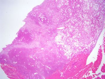
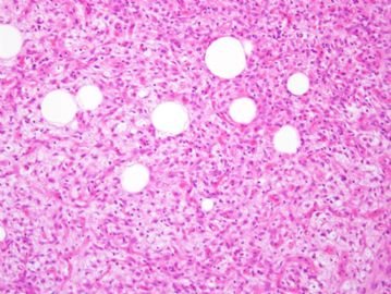
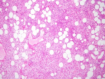
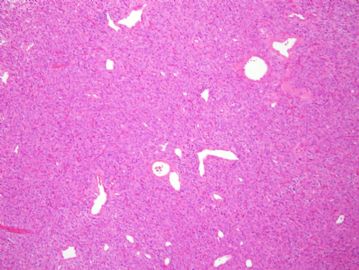
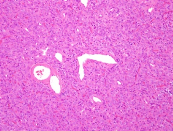
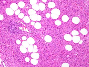
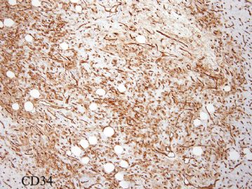
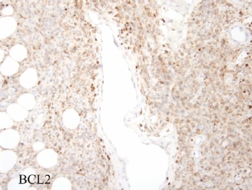
诊断: 脂肪瘤样SFT/HPC lipomatous solitary fibrous tumor/hemangiopericytoma
下面这篇文献做过超微结构证明 lipomatous hemangiopericytoma 具有SFT的结构,所以看作是SFT的一个型。
Hum Pathol. 2000 Sep;31(9):1108-15.
Lipomatous hemangiopericytoma: a fat-containing variant of solitary fibrous tumor? Clinicopathologic, immunohistochemical, and ultrastructural analysis of a series in favor of a unifying concept.
Guillou L, Gebhard S, Coindre JM.
University Institute of Pathology, Lausanne, Switzerland.
The dinicopathologic, immunohistochemical and ultrastructural features of 13 lipomatous hemangiopericytomas are presented. There were 6 male and 7 female patients whose ages at diagnosis ranged from 27 to 75 years (median 48) all presenting with a mass of variable duration. The tumor sizes ranged from 1.7 cm to 19 cm (median 5.5 cm). The locations included the orbit (1), neck (1), mediastinum (1), epicardium (1), retroperitoneum (3), right iliac fossa (1), and upper (1) and lower (4) extremity. Histologically, the lesions were composed of a varying admixture of spindle-shaped to round cells, variably collagenous stroma, adipose tissue, and branched, often thick-walled, hemangiopericytoma-like vessels. For 11 tumors, the mitotic activity ranged from 1 to 3 mitoses per 10 high-power fields (HPF). One tumor which contained hypercellular areas showed 13 mitoses per 10 HPF, and another hypercellular lesion showed up to 43 mitoses per 10 HPF, abnormal mitoses, and necrosis. Immunohistochemically, tumor cells were invariably positive for vimentin and CD99, and mostly for CD34 but negative for desmin, keratin, CD31, CD117 (c-kit), and inhibin. About half of the tumors showed reactivity for bcl-2. Occasionally, focal reactivity was also observed for smooth muscle actin, muscle-specific actin, S100 protein, and epithelial membrane antigen. Ultrastructural examination of seven cases showed features in keeping with fibroblastic, myofibroblastic, or pericytic differentiation. Treatment consisted of simple tumorectomy in 10 cases and wide excision in 3. Follow-up information on 10 patients (range: 6 to 77 months; median: 18 months) showed no recurrence. Lipomatous hemangiopericytoma which share the clinical, pathologic, immunohistochemical, and ultrastructural features of solitary fibrous tumor (SFT) is likely to represent, in most cases, a fat-containing variant of SFT.
-
wangdingding 离线
- 帖子:1474
- 粉蓝豆:98
- 经验:6042
- 注册时间:2006-10-19
- 加关注 | 发消息

