| 图片: | |
|---|---|
| 名称: | |
| 描述: | |
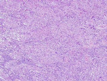
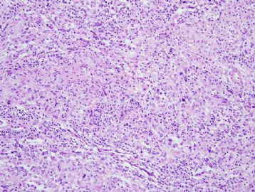
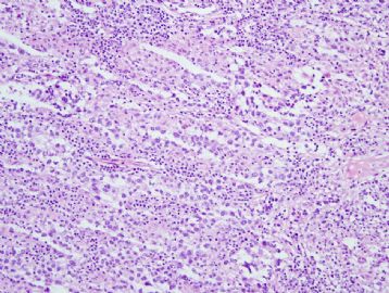
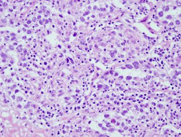
- 60 year old man with mediastinal mass
-
stevenshen 离线
- 帖子:343
- 粉蓝豆:2
- 经验:343
- 注册时间:2008-06-03
- 加关注 | 发消息
-
stevenshen 离线
- 帖子:343
- 粉蓝豆:2
- 经验:343
- 注册时间:2008-06-03
- 加关注 | 发消息
-
本帖最后由 于 2010-03-08 10:05:00 编辑
Thank you for asking me to stick my neck out in public. Risky as it is, I am old and brave enough to be embarrassed and possibly humiliated for the sake of discussion and education.
Mediastinal or, more correctly, ANTERIOR mediastinal mass has the proverbial 3 Ts in differential diagnoses - Thymic neoplasms, Thyroid neoplasms, and Teratoma and related neoplasms. There are no features of thymolipoma, hyperplastic thymus, non-Hodgkin lymphoma, papillary carcinoma of thymus, carcinoid or neuroendocrine tumors, thyroid follicular or papillary neoplasms, spindle epithelial tumor with thymus-like element, or mature teratomatous elements. Instead, there are many epithelioid granulomas in the background and irregular aggregates of large neoplastic cells with vesicular nuclei, large prominent nucleoli, vacuolated cytoplasm in association with many infiltrating small lymphocytes. Individual apoptotic bodies are found among the large neoplastic cells, suggestive of malignancy. Thymoma and thymic carcinoma may have intimately associated small lymphocytes (thymocytes), but their epithelial elements are either organoid and cohesive nests of epithelial cells with oval or elongated nuclei, or frankly malignant in architecture and cytology. While epithelioid granulomas are seen in rare thymic neoplasms, they are a frequent finding in seminoma, dysgerminoma and germinoma, all of which invariably show varying degrees of small lymphocytic infiltration. That's how I reached my conclusion.
At intraoperative consultation, we are pressured to provide a confident diagnostic interpretation to the surgeons, most of whom do not have the patience to listen to a list of differential diagnoses of longer than 2. Therefore I have always tried to give them only one diagnosis whenever I can. This approach may result in occasional mistakes, so it has to be taken very carefully or else we (and, more critically, the surgeons) quickly lose confidence in our important role as intraoperative consultants. Are there important differential diagnoses in my consideration of this case? Sure there were. Classic Hodgkin lymphoma is also known to be granulomatous on occasion, and Reed-Sternberg cells and its variants may rarely appear very epithelioid in large aggregates. Malignant T cells seen in anaplastic large cell lymphomas often are sinusoidal in distribution and may appear very epithlioid. Metastatic carcinoma and melanoma in lymph nodes may mimic anything. Lastly, metastatic malignant mesothelioma and benign mesothelial displacement into lymph nodal sinusoids may appear similarly. Permanent sections with better preservation of architectural and cytologic details of the entire specimen certainly will provide us with more confidence in what we believe this is, and immunohistochemistry is essential for its confirmation. That said, I don't think this case is any of these differential diagnoses mentioned.

聞道有先後,術業有專攻
-
stevenshen 离线
- 帖子:343
- 粉蓝豆:2
- 经验:343
- 注册时间:2008-06-03
- 加关注 | 发消息
- Thanks for Dr. Ma's great comments and summary of a very logic diagnostic process to a correct diagnosis.
- It is well known diagnostic pitfalls for seminoma (dysgerminoma) when there is extensive granulomatous inflammation in gonadal or extra-gonadal locations.
- I also enjoy reading Dr. Ma comment about the principles of frozen diagnosis. This confidence has to be build upon solid knowledge and diagnostic skills, as well as many years of experience.


