| 图片: | |
|---|---|
| 名称: | |
| 描述: | |
- 请求会诊——初步诊断——间变型PXA
-
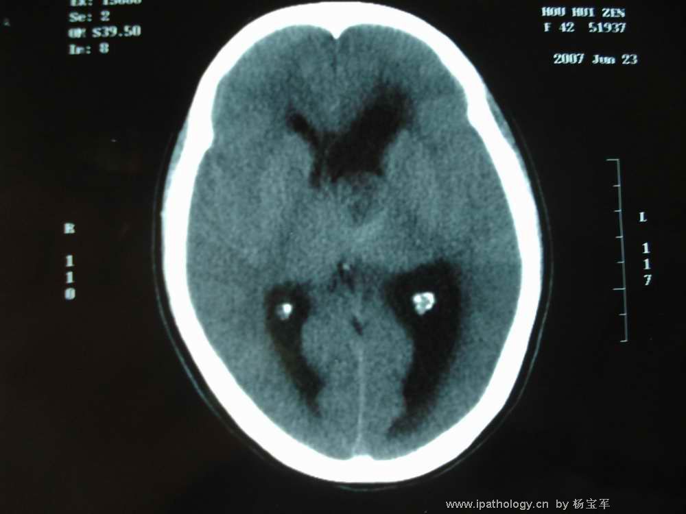
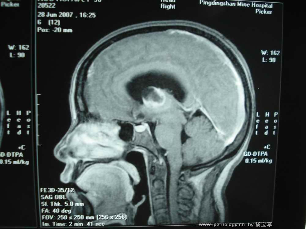
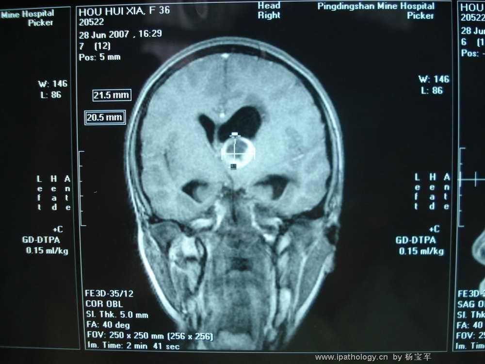
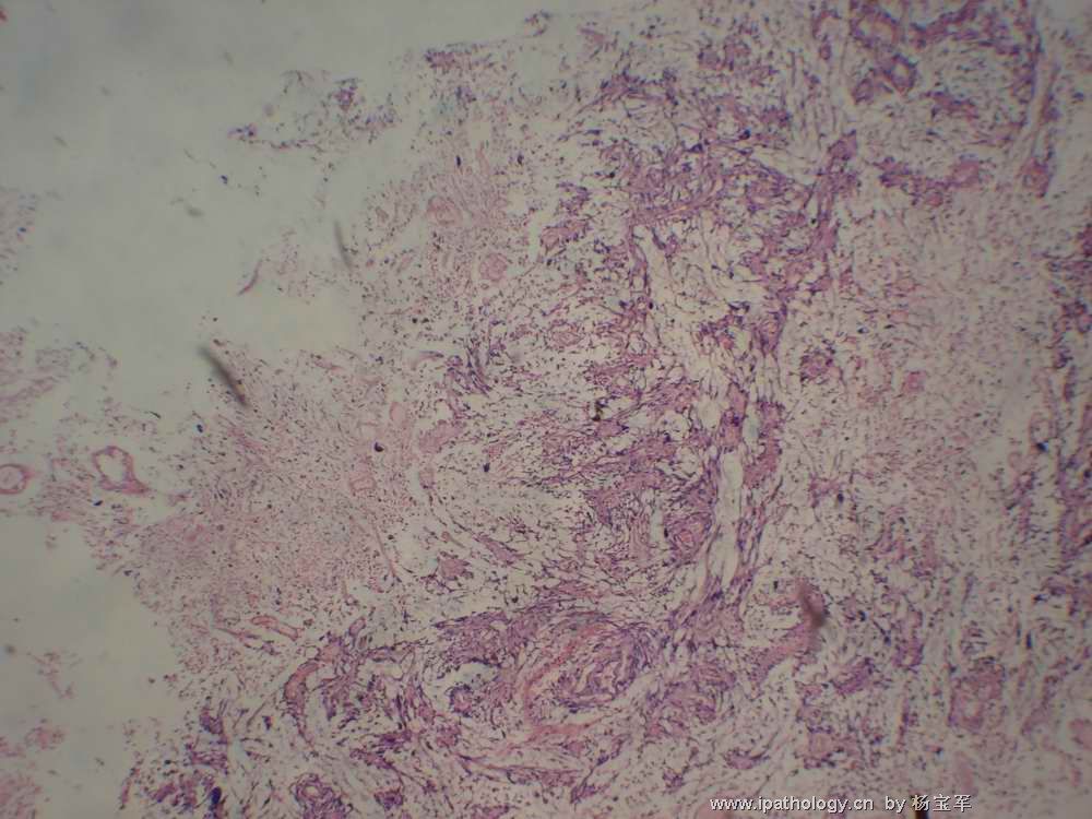
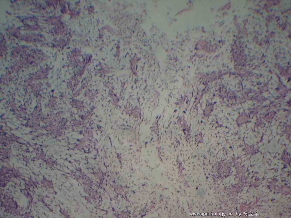
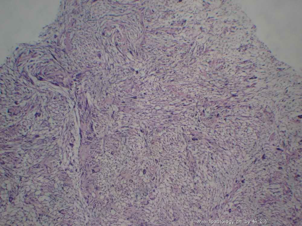
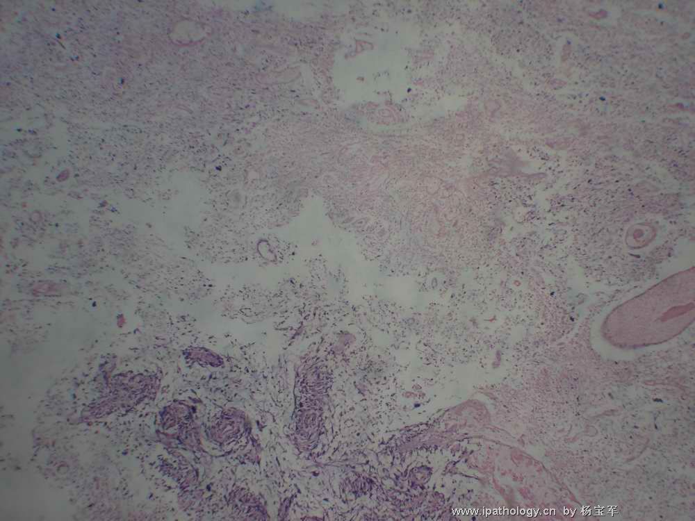
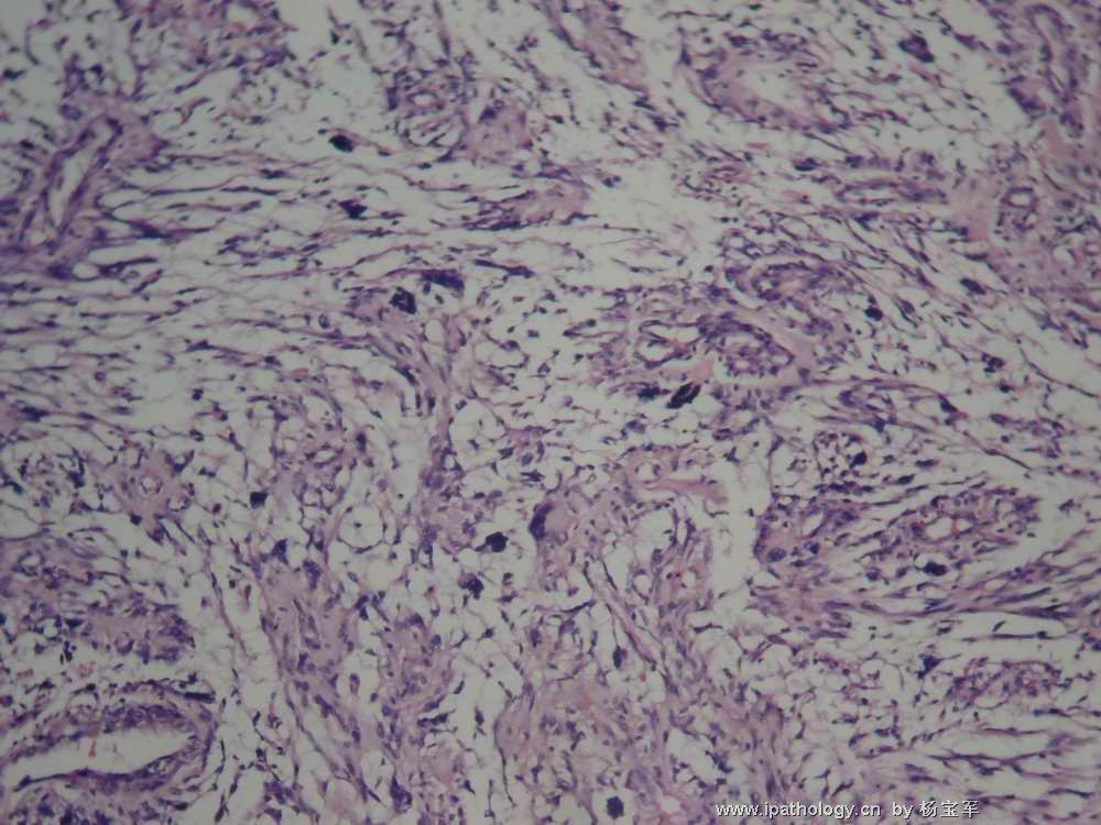
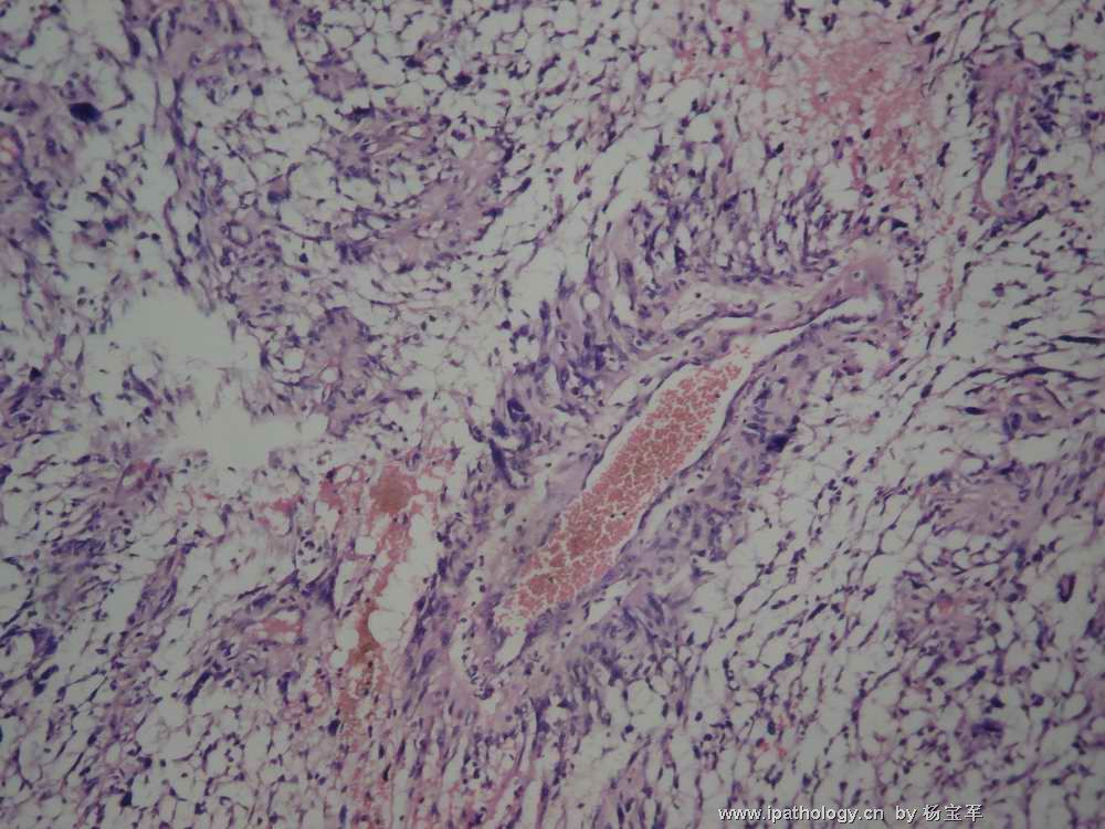
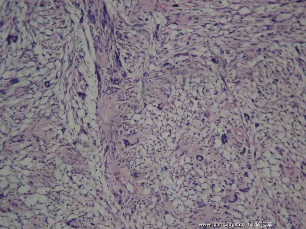
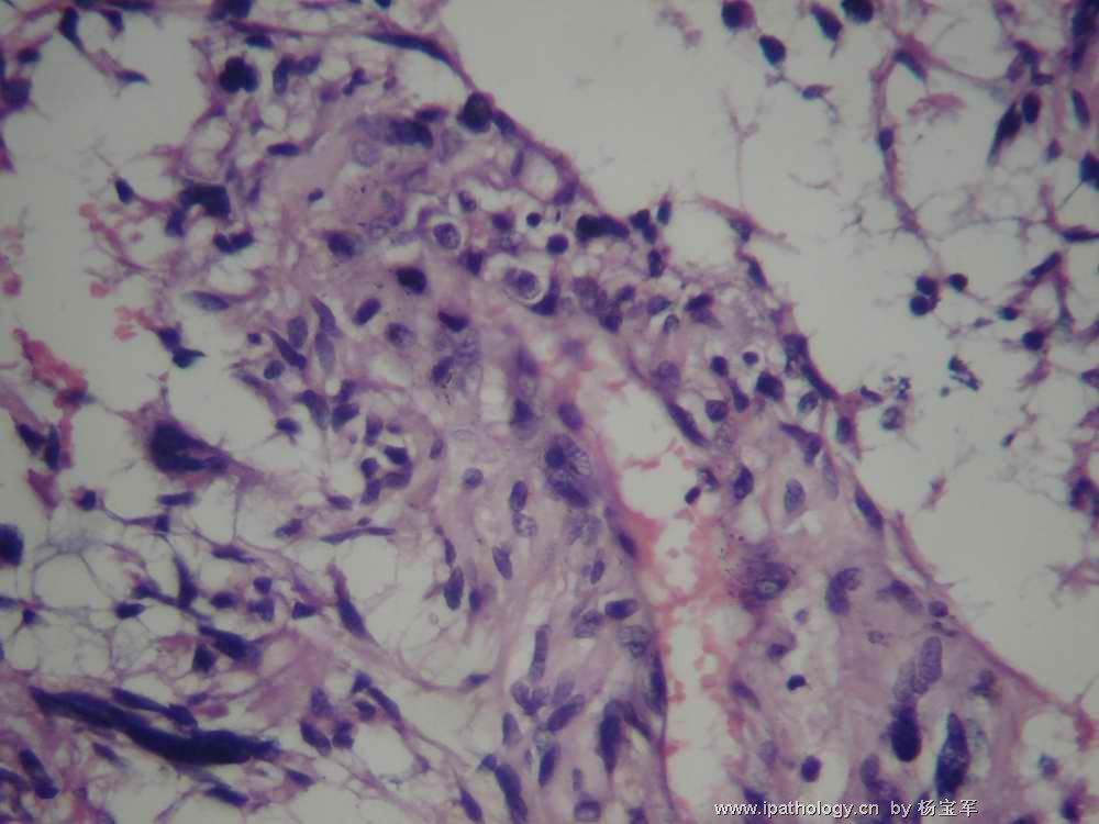
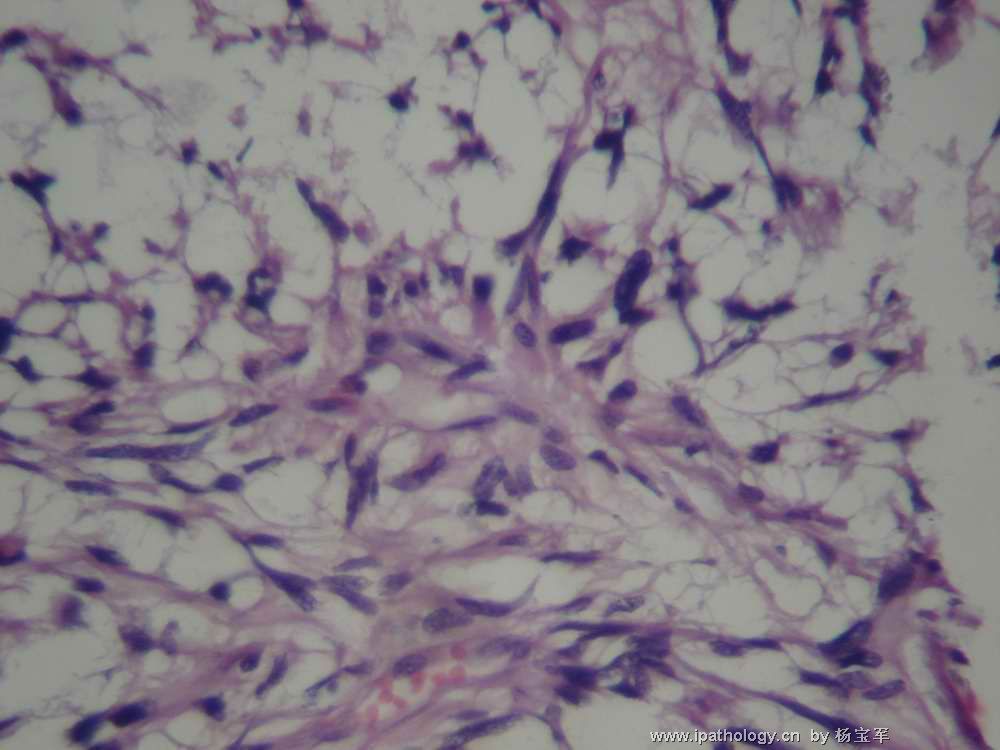
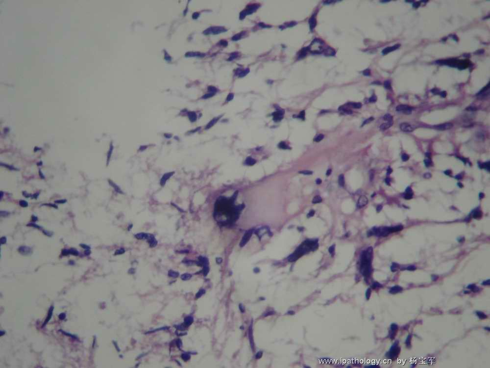
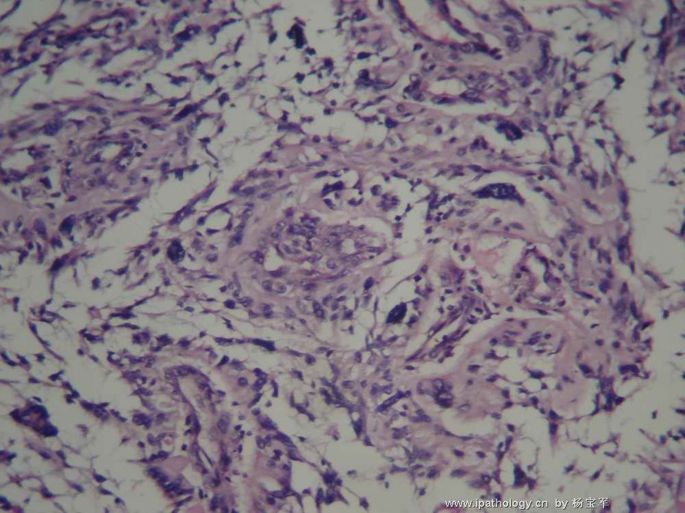
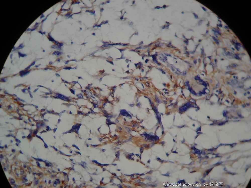
I think this is a glioblastoma, WHO grade IV, because of glial differentiation, marked cytologic atypia, prominent tumor mecrosis, and focal possible vascular proliferation. Though I do not see obvious mitotic figures in the photos, I don't doubt there are some. Certainly, the relatively young patient's age and the circumscription of the lesion on MRI and CT scans suggest less malignant lesions like PXA. However, there are no eosinophilic granular bodies or characteristic perivascular inflammation to support PXA.

聞道有先後,術業有專攻
-
本帖最后由 于 2007-07-11 12:38:00 编辑
I think this is a glioblastoma, WHO grade IV, because of glial differentiation, marked cytologic atypia, prominent tumor mecrosis, and focal possible vascular proliferation. 由于有胶质分化,细胞异形性以及明显的肿瘤性坏死和局灶性血管增生,我考虑是胶质母细胞瘤,WHOIV级, Though I do not see obvious mitotic figures in the photos, I don't doubt there are some. 提供的图像中没有看到明显的核丝分裂,我认为肯定有一些核丝分裂.Certainly, the relatively young patient's age and the circumsc ription of the lesion on MRI and CT scans suggest less malignant lesions like PXA. However, there are no eosinophilic granular bodies or characteristic perivascular inflammation to support PXA. 尽管患者年龄较青,MRI和CT提示为低度恶性PXA样病变,但是没有嗜酸性颗粒小体、没有特征性的血管周围炎症,不支持PXA。

