| 图片: | |
|---|---|
| 名称: | |
| 描述: | |
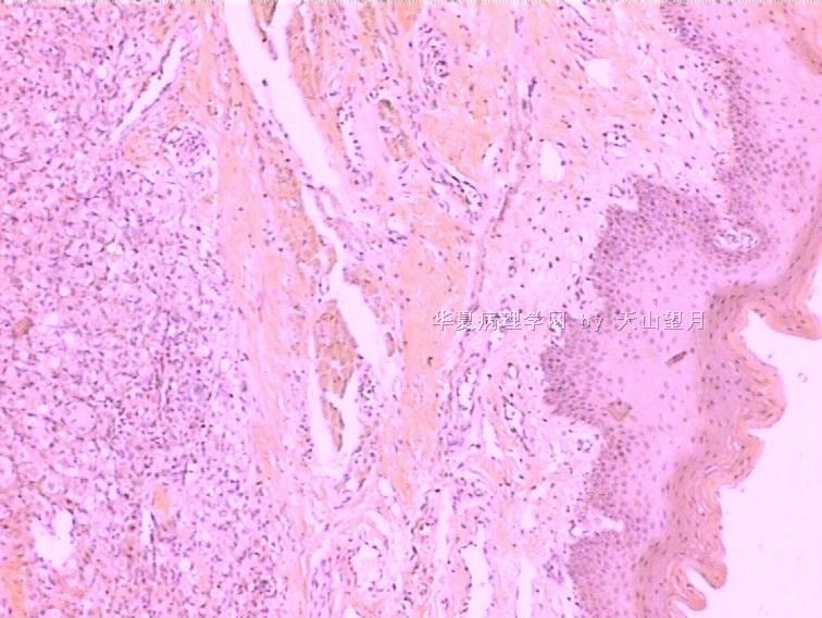
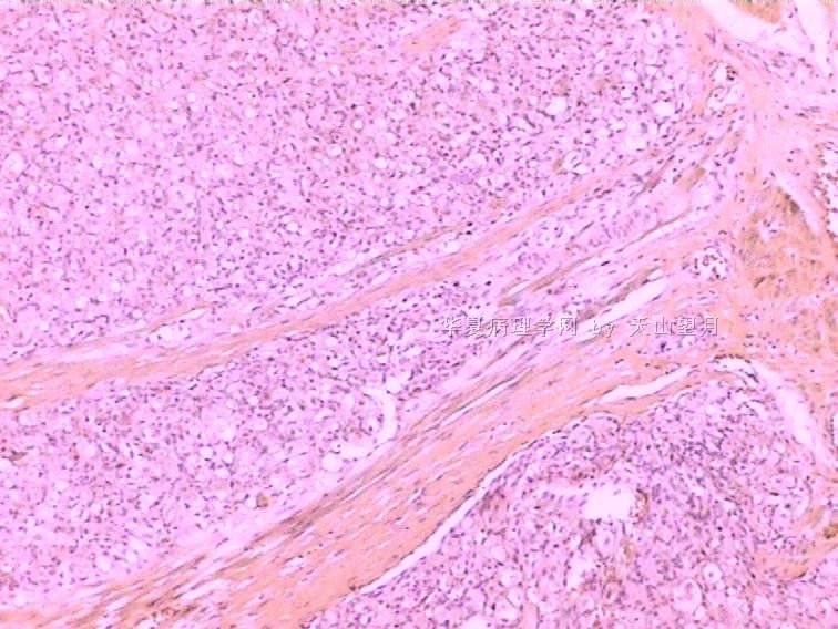
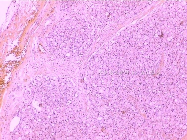
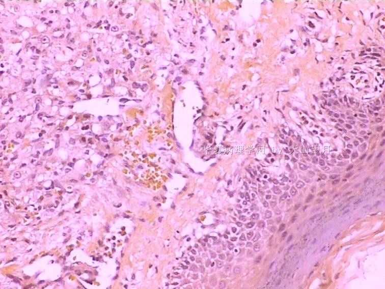
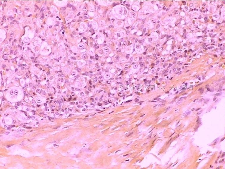
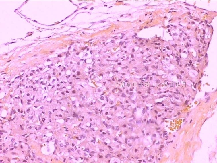
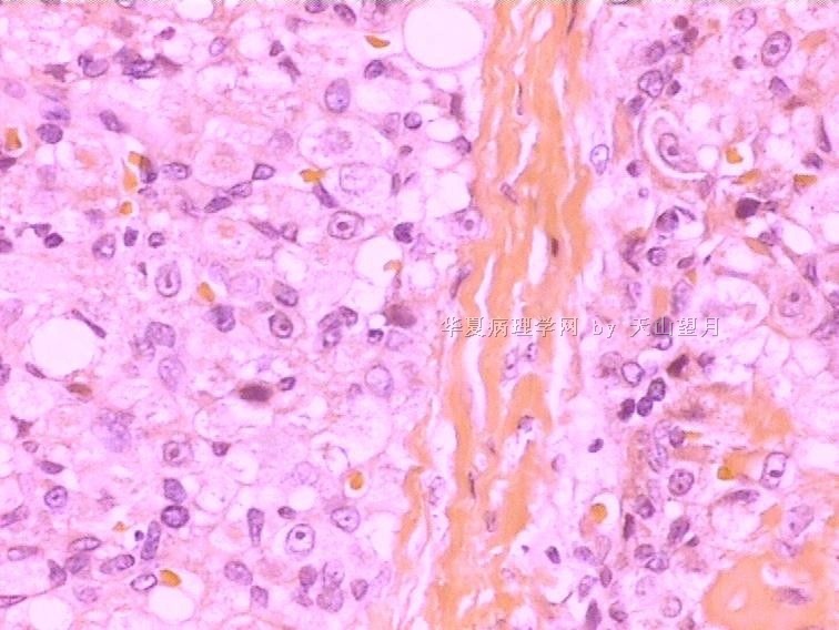
- 4岁,男孩,舌头结节。
-
本帖最后由 于 2010-02-24 22:55:00 编辑
Although many cells between vacuoles and collapsed capillaries/sinuses appear epithelioid with oval vesicular nuclei, single nucleoli and plump cytoplasm, I do not see enough atypia to worry about malignant neoplasm. The lobular architecture, the anatomic location (underneath tongue mucosa) and numerous associated blood vessels are consistent with that of a lobular capillary hemangioma (or pyogenic granuloma), but the epithelioid appearance of constituent cells would suggest epithelioid hemangioendothelioma.

聞道有先後,術業有專攻
-
本帖最后由 于 2010-02-24 23:01:00 编辑
纤维分隔小叶状,小叶中2种细胞,一种圆形或卵圆形,核圆形,空淡,核仁明显,一种胞浆透亮,核印戒状,包绕前者,形成小巢状。可见丰富的血管、裂隙、色素(含铁血黄色?黑色素?其他?)、散在红细胞。考虑
1、血管源性肿瘤:上皮样血管瘤?上皮样血管内皮瘤?血管肉瘤?
2、血管母细胞瘤?
3、幼年型色素痣?不知舌头能发生吗?
4、恶黑?年龄太小。
5、纤维组织细胞瘤?硬化性血管瘤?
6、颗粒细胞瘤?舌头容易发生,但此例不像。
呵呵,范老师提供的,一定难度大,我先猜猜吧。大家继续,谢谢!

- 广州金域病理
-
1212121212 离线
- 帖子:37
- 粉蓝豆:1
- 经验:37
- 注册时间:2009-12-06
- 加关注 | 发消息
| 以下是引用mjma在2010-2-24 22:53:00的发言: Although many cells between vacuoles and collapsed capillaries/sinuses appear epithelioid with oval vesicular nuclei, single nucleoli and plump cytoplasm, I do not see enough atypia to worry about malignant neoplasm. The lobular architecture, the anatomic location (underneath tongue mucosa) and numerous associated blood vessels are consistent with that of a lobular capillary hemangioma (or pyogenic granuloma), but the epithelioid appearance of constituent cells would suggest epithelioid hemangioendothelioma. |



 ,我还是等答案吧


