| 图片: | |
|---|---|
| 名称: | |
| 描述: | |
- (已确诊)右卵巢肿块(3)
请见下文卵巢妊娠黄体瘤伴有颗粒细胞增生---酷似卵巢(恶性)肿瘤的描述,
Pathol Res Pract. 1999;195(12):859-63.
Pregnancy luteoma with granulosa cell proliferation: an unusual hyperplastic lesion arising in pregnancy and mimicking an ovarian neoplasia.
Piana S, Nogales FF, Corrado S, Cardinale L, Gusolfino D, Rivasi F.
Department of Morphological Sciences and Forensic Medicine, University of Modena and Reggio Emilia, Italy.
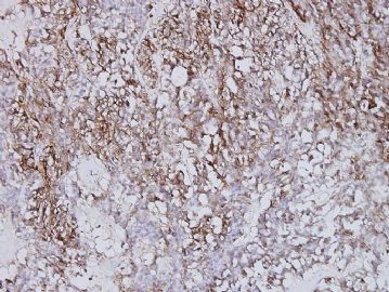
A pregnancy luteoma (PL) was incidentally found at a term cesarean section in a 27-year-old black woman without any endocrine abnormality. The lesion involved only the left ovary; it had a nodular and focal pseudoalveolar growth pattern and was associated with areas of tubular sertoliform component, consistent with granulosa cell proliferation. Immunohistochemistry revealed a diffuse positivity to Inhibin A, CD99, cytokeratin and vimentin. The ultrastructure was typical of steroid-producing cells. PL is a tumor-like lesion arising in pregnant women and often misdiagnosed as a neoplastic lesion; awareness of this rare entity and its differential diagnoses may avoid unnecessary surgery in young patients.

- 王军臣
-
本帖最后由 于 2010-02-23 17:49:00 编辑
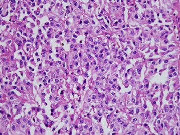
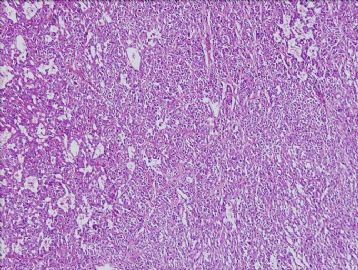
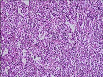
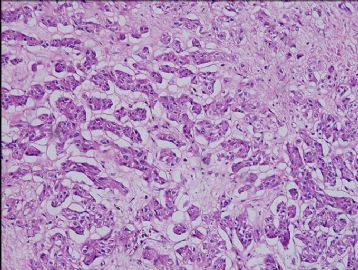
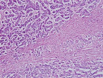
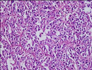
1.病史:孕4月,卵巢肿块。
2.大体:肿块大小2x1.3x1.1cm,灰白黄色。
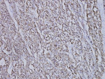
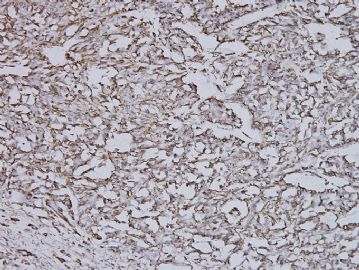
3.组织学:细胞为多边形或不规则形,构成内分泌样结构。胞浆丰富、淡染或有的比较透明,核异型性不明显。在高倍下,细胞学形态有的类似于黄素化的间质细胞,有的像肾上腺皮质细胞,有的酷似黄体细胞。多数细胞排成密集的腺巢状;有的呈梁状或条索状,局部间质粘液样水肿。
4.诊断:妊娠黄体瘤。结合病史,这一诊断的可能性最大。
5.鉴别诊断:性索-间质肿瘤(支持细胞-间质细胞肿瘤),主要鉴别 Leydig cell tumor(支持细胞瘤)。
妊娠性黄体瘤实际上是增生性病变(瘤样病变),而非真正的肿瘤

- 王军臣

