| 图片: | |
|---|---|
| 名称: | |
| 描述: | |
- 移植肾穿刺病例
-
本帖最后由 于 2010-02-06 10:49:00 编辑
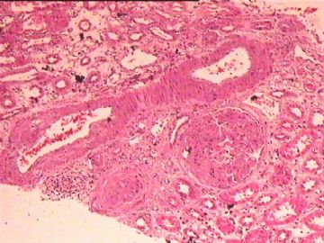
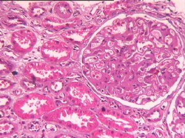
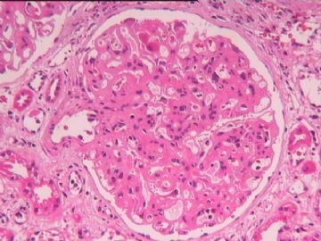
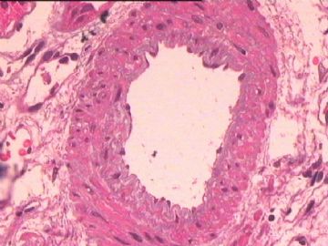
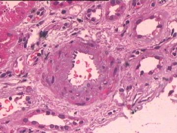
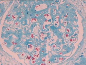
The glomerular basement membrane appears thickened and the mesangia are expanded. Without silver stain, it is difficult to appreciat the double contour. If the double contour is truely present, chronic transplant glomerulopathy is the diagnosis. The artery shows intimal thickening, suspicious for chronic transplant arteriopathy. But the trichrome stain (photo 5?) fails to demonstrate neo-media formation.
-
本帖最后由 于 2010-02-09 07:32:00 编辑
I agree with Dr. Frankbj's observation of this biopsy (cg3, ct2, ct2,cv1,mm3). But I have a different interpretation of cg3,ci2,ct2,cv1,mm3.
1. cg3: it is defined as double contours affecting more than 50% of peripheral capillary loops in the most affected of nonsclerotic glomeruli. When we see cg3, it is considered as chronic transplant glomerulopathy, part of chronic active antibody-mediated rejection. Positive C4d is NOT required for this diagnosis.
2. ci2, ct2: ci2 is defined as interstitial fibrosis of 26 to 50% of cortical area; ct2 is defined as tubular atrophy involving 26 to 50% of the area of cortical tubules. These changes are non-specific. There are many factors which may cause ci and ct.
3.cv1: it is defined as vascular narrowing of up to 25% lumenal area by fibrointimal thickening of arteries ± breach of internal elastic lamina or presence of foam cells or occasional mononuclear cells. Fibrointimal thickening itself is not regarded as specific for chronic active T-cell-mediated rejection. If we saw breach of internal elastic lamina or presence of foam cells or occasional mononuclear cells, these were considered as "chronic rejection" in the past. In 2007 update, chronic active T-cell-mediated rejection is defined as presence of ‘chronic allograft arteriopathy’ (arterial intimal fibrosis with mononuclear cell infiltration in fibrosis, formation of neo-intima).
4. mm3: mm is defined as the expanded mesangial interspace between adjacent capillaries. If >50% of nonsclerotic glomeruli affected (at least moderate matrix increase), it is defined as mm3. mm was considered as a potentially importan but less specific finding for chronic transplant glomerulopathy. If I recall correctly, in 2005 Banff meeting, a young pathologist suggested to remove mm from Banff schema. That attempt failed.
Therefore, (cg3,ci2,ct2,cv1,mm3) should be interpreted as combined chronic active antibody-mediated rejection and probable chronic active T-cell-mediated rejection.

