| 图片: | |
|---|---|
| 名称: | |
| 描述: | |
- 腹部肿物
-
本帖最后由 于 2010-01-29 22:35:00 编辑
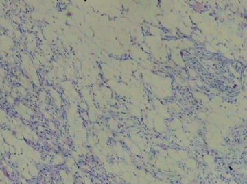
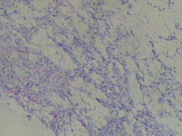
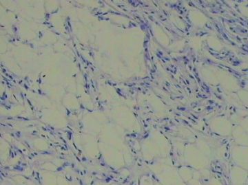
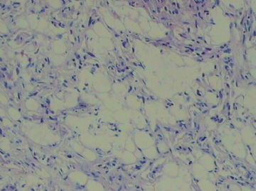
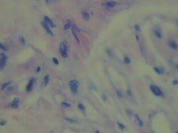
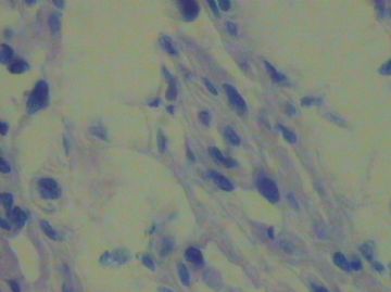
The photos suggest spindle cell lipoma, which often co-exists with atypical lipomatous tumor or well differentiated liposarcoma depending on the anatomic location of the tumor. The small size of this tumor (1 cm) makes this unlikely, but sometimes retroperitoneal liposarcoma looks just like fat grossly. Can you make sure that the tumor is from abdominal wall (subcutaneous tissue) and not from mesentery or deep soft tissue? If it is from the retroperitoneum, one has to look carefully for features atypical lipomatous tumor/liposarcoma.

聞道有先後,術業有專攻
