| 图片: | |
|---|---|
| 名称: | |
| 描述: | |
- 宫颈液基
| 姓 名: | ××× | 性别: | 女 | 年龄: | 44 |
| 标本名称: | 宫颈赘生物 | ||||
| 简要病史: | 体检,无临床症状 | ||||
| 肉眼检查: | 体积:0.3x0.2x0.1 | ||||
-
本帖最后由 于 2010-01-01 10:44:00 编辑

- Stop walking today and you'll have to run tomorrow.
| 以下是引用mingfuyu在2010-1-2 23:32:00的发言:
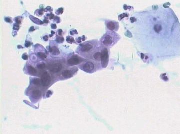
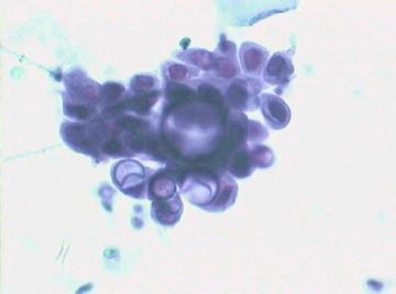
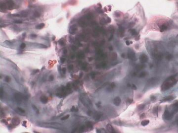
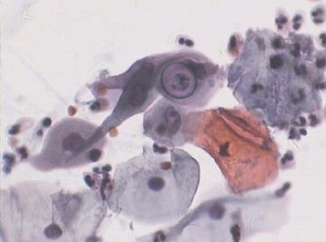
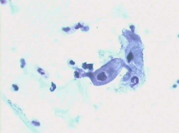
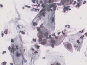
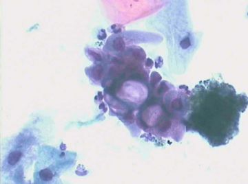
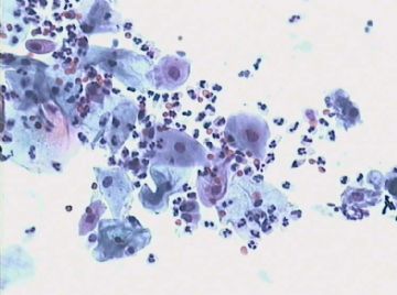
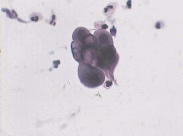
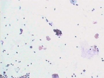
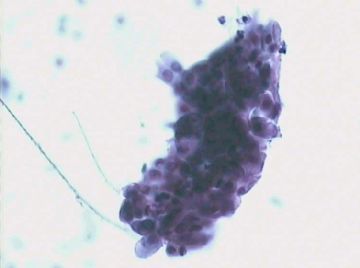
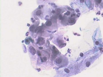
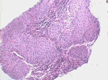
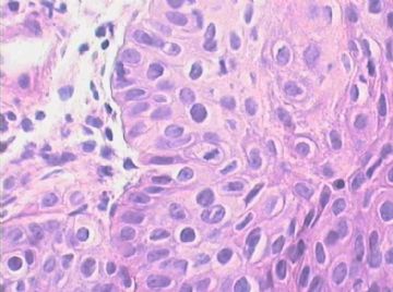
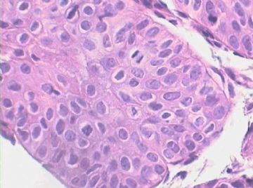
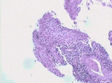
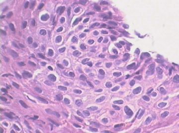
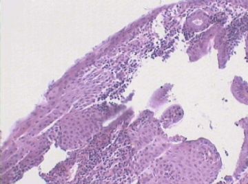
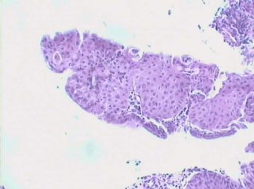
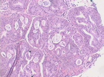
Pap: my impression is NILM. Most "worrisome" cells are endocervical glandular cells and metaplastic cells. No cells show enlarged nuclei more than 2.5X the size of normal intermediate cells, very fine and even chromatin and very smooth nuclear membrane. The bubble-like spaces are probably from cytoplasmic mucin of endocervical glandular cells. Absolutely no typical HPV cytopathic changes, so-called koilocytes. Of course some HPV infected cells don't show koilocytic changes but nuclear atypia must be present to diagnose or suspect HPV infection. Biopsy; No difinite CIN. Squamous metaplasia present with some mitoses which is acceptible at the lowr portion of the epithelium for reactive/reparative changes. As seen in the pap, nuclei are very uniform between cells, fine and even chromatin and very smooth nuclear membrane. Absolutely no typical HPV associated changes. The maxmum interpretation you can do is atypical metaplasia, then do p16 to rule out CIN2. If the shown photos are the worst area, in real work, i would sign out as negative for dysplasia. It is nice to show the pap and biopsy together. They correlate with each other and both support negative for dysplasia. |
巴氏片:我认为是NILM。多数“麻烦”细胞是子宫内膜腺细胞和化生细胞,细胞胞核增大未超过正常中层细胞核的2.5X,染色质细腻均匀、核膜非常平滑,泡沫样间隙可能来源于子宫内膜腺细胞的胞质内粘液。完全未见HPV效应所致的所谓“空穴细胞”,当然部分HPV感染细胞也可能不表现为“空穴细胞”改变,但其胞核的非典型性应该可以提示或怀疑HPV感染。
活检片:无明确CIN病变。上皮底层反应性/修复性改变所致的有部分有丝分裂活性的鳞化是可以接受的。正如巴氏片中所见,细胞的胞核非常一致、染色质细腻均匀、核膜平滑,无典型HPV相关改变。最多判读为:不典型化生,然后做p16以除外CIN2可能。如果所示图片是病变最重的区域,实际工作中,我会签发阴性发育不良报告。
将巴氏片和活检片一起展示,很好,二者相互关联并且都支持阴性发育不良的判读。
多谢喻老师来指导!青青子衿
Pap: my impression is NILM. Most "worrisome" cells are endocervical glandular cells and metaplastic cells. No cells show enlarged nuclei more than 2.5X the size of normal intermediate cells, very fine and even chromatin and very smooth nuclear membrane. The bubble-like spaces are probably from cytoplasmic mucin of endocervical glandular cells. Absolutely no typical HPV cytopathic changes, so-called koilocytes. Of course some HPV infected cells don't show koilocytic changes but nuclear atypia must be present to diagnose or suspect HPV infection.
Biopsy; No difinite CIN. Squamous metaplasia present with some mitoses which is acceptible at the lowr portion of the epithelium for reactive/reparative changes. As seen in the pap, nuclei are very uniform between cells, fine and even chromatin and very smooth nuclear membrane. Absolutely no typical HPV associated changes. The maxmum interpretation you can do is atypical metaplasia, then do p16 to rule out CIN2. If the shown photos are the worst area, in real work, i would sign out as negative for dysplasia.
It is nice to show the pap and biopsy together. They correlate with each other and both support negative for dysplasia.

