| 图片: | |
|---|---|
| 名称: | |
| 描述: | |
- B1-9(天津医科大学附属肿瘤医院)_已点评
-
zhaoyan2006 离线
- 帖子:139
- 粉蓝豆:42
- 经验:301
- 注册时间:2009-02-23
- 加关注 | 发消息
REVIEW
Recent developments in the histological diagnosis of
spindle cell carcinoma, fibromatosis and phyllodes tumour
of the breast
A H S Lee
Histopathology Department, Nottingham University Hospitals, City Hospital Campus, Nottingham, UK
This article reviews recent advances in the diagnosis of
these three unusual tumours of the breast. Spindle cell
carcinoma needs to be considered in the differential
diagnosis of many mammary spindle cell lesions: it is
important to be aware of the wide range of appear-
ances, including the recently described fibromatosis-
like variant. Immunohistochemistry using a broad
panel of cytokeratin antibodies is needed to exclude
spindle cell carcinoma; there is frequent expression of
basal cytokeratins and p63. CD34 is often expressed by
the stroma of phyllodes tumours, but does not appear
to be expressed by spindle cell carcinoma or fibroma-
tosis. Nuclear b-catenin is found in about 80% of
fibromatoses, but can also be seen in spindle cell
carcinomas and phyllodes tumours. Two recent studies
have described features useful in the distinction of
phyllodes tumour and fibroadenoma on core biopsy,
including increased cellularity, mitoses and overgrowth
of the stroma, adipose tissue in the stroma and
fragmentation of the biopsy specimen. Periductal stro-
mal tumour is a recently described biphasic tumour
composed of spindle cells around open tubules or ducts
(but no leaf-like architecture) with frequent CD34
expression. The overlap of morphology with phyllodes
tumour suggests that it may be best regarded as a
variant of phyllodes tumour.
读片会讨论意见:
炎症性肌纤维母细胞肿瘤
肉瘤,叶状囊肉瘤或恶性叶状瘤
纤维瘤病样化生性癌
(右乳)化生性癌(梭形细胞癌)
(右乳)分叶状肿瘤,核分裂象2-5、10HPF,交界性
化生性癌-梭形细胞癌
化生性癌
Metaplastic carcinoma
右乳)梭形细胞癌。做免疫组化排除恶性叶状肿瘤(低级别)、低级别肉瘤或增生活跃的瘤样病变
炎症性肌纤维母细胞瘤,化生性癌待排
化生性癌
出片单位意见:梭形细胞癌
免疫组化结果:CK(+)
华夏病理/粉蓝医疗
为基层医院病理科提供全面解决方案,
努力让人人享有便捷准确可靠的病理诊断服务。
-
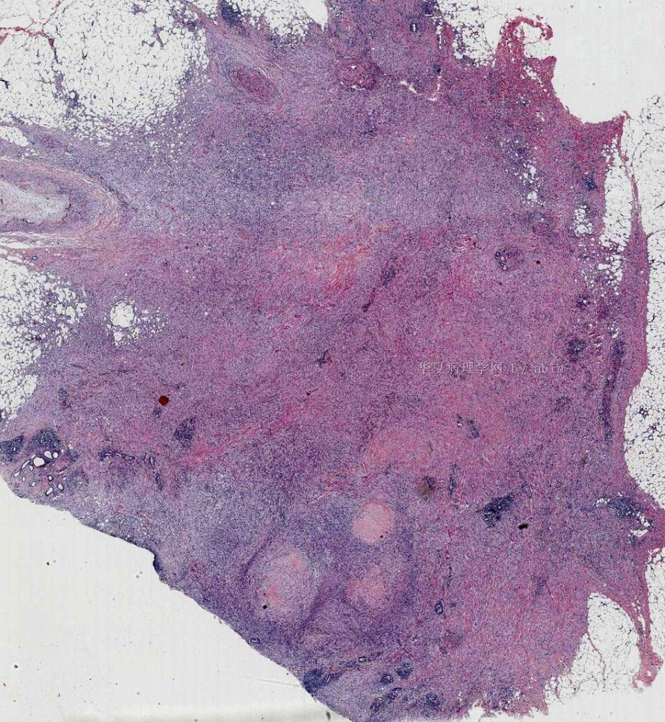
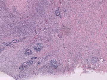
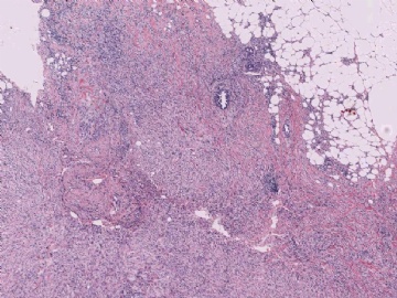
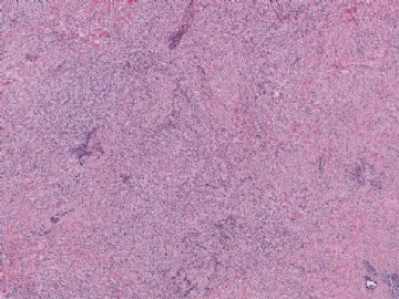
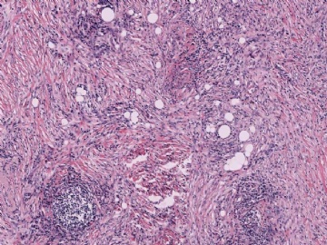
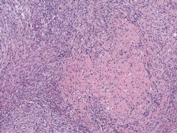
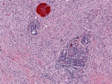
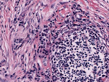
这一例从形态学上我比较赞同大家的诊断,即所谓的低度恶性的纤维瘤病样的梭形细胞癌。将这个诊断与一般的化生性癌或梭形细胞癌区分开还是有实际价值的,因为通常临床医生会认为化生性癌的预后不佳,而低度恶性的纤维瘤病样的梭形细胞癌是一种低度恶性的肿瘤,不同于大多数化生性癌或三阴性乳腺癌,有些文章中也不主张进行腋窝淋巴结清扫或术后辅助化疗。下面是我曾经写过的一篇关于该肿瘤介绍中的部分内容。
纤维瘤病样梭形细胞癌的诊断标准如下:(1)肿瘤主要(≥95%)由梭形细胞构成,细胞有轻-中度异型;(2)浸润性上皮成分不超过肿瘤的5%,且浸润性成分不位于周边。
纤维瘤病样梭形细胞癌可进一步分为单相型和双相型2种类型。单相型肿瘤中缺乏明显的上皮成分,但可见上皮样细胞团。这种“上皮样细胞团”是其它梭形细胞病变中所不具备的。双相型肿瘤形态学上可见灶性的浸润性癌区域,或出现肿瘤性的鳞状细胞,这些细胞可与梭形细胞成分相移行。
肿瘤细胞以梭形/纤维母细胞样或星形/肌纤维母细胞样细胞增生为主,呈编织状或束状排列,瘤细胞间常有多少不等的胶原。瘤细胞形态温和或有轻-中度异型,核分裂象少见(<2个/10HPF),少数可达5个/10HPF。除梭形细胞成分外,肿瘤内还可见散在上皮成分(
一般<5%),多为鳞状上皮或腺上皮伴轻度异型,或仅表现为梭形或多边形细胞有上皮样分化。
免疫表型:梭形细胞常成片表达Vimentin,CK34B12,
CKAE1/3,仅单个细胞阳性少见,而低分子量角蛋白CK7则较少表达,EMA部分弱表达,SMA主要在CK阴性细胞表达,少数与CK阳性细胞共表达。这提醒我们在选择上皮标志物时应选择广谱CK或高分子量CK,而不要选择低分子量CK。该肿瘤为三阴性乳腺癌,不表达ER,PR,Her2/neu。
Sneige等对24例乳腺低度恶性纤维瘤病样梭形细胞癌的研究中,虽未发现腋窝淋巴结转移者,但2例单纯肿块切除者有局部复发,另2例改良根治术患者于2年内发生肺转移并最终死亡,肺转移灶形态同乳腺原发灶,有明显的纤维化和胶原。此2例形态上呈明显纤维瘤病样表现,核分裂象少(<1个/10HPF),但肿瘤较大(最大径分别为

