| 图片: | |
|---|---|
| 名称: | |
| 描述: | |
- 骶髂肿物
| 姓 名: | ××× | 性别: | 年龄: | ||
| 标本名称: | |||||
| 简要病史: | |||||
| 肉眼检查: | |||||
女性,21岁。骶髂肿物10cmx10cm大小边界不清,侵犯脊骨组织和周围组织。
免疫组化:
CK、CD99、 Desmin 、S-100 、SMA VIM均-
NSE散在+ BCL-2散在+
CD34血管+ SMA血管+
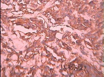
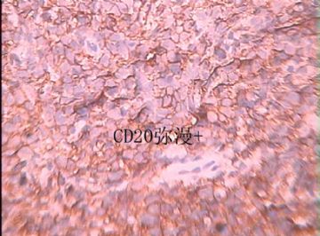
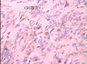
LCA弥漫+ CD20弥漫+ CD3散在+
-
本帖最后由 于 2009-12-14 18:21:00 编辑
Mod Pathol. 2001 Nov;14(11):1175-82.
Differentiating lymphoblastic lymphoma and Ewing's sarcoma: lymphocyte markers and gene rearrangement.
Ozdemirli M, Fanburg-Smith JC, Hartmann DP, Azumi N, Miettinen M.
Department of Pathology, Georgetown University Medical Center, 3900 Reservoir Road N.W., Washington, DC 20007, USA.
We encountered a child with an intraosseous small round cell tumor that was negative for LCA, CD20 (L26), and CD3 and positive for vimentin, CD99 (MIC-2), and periodic acid-Schiff. The tumor exhibited rosette-like formations. This case was initially interpreted as Ewing's sarcoma (ES); however, additional studies revealed positivity for CD79a, CD43, and TdT expression, and an immunoglobulin heavy chain gene rearrangement (IgH-R) by polymerase chain reaction (PCR) established this to be a precursor B-lymphoblastic lymphoma. Because the differential diagnosis of ES and lymphoblastic lymphoma can be difficult and the differential diagnostic value of leukocyte antigens and immunoglobulin heavy chain gene rearrangement studies have not been fully evaluated, we conducted a more extensive investigation on 33 (21 soft tissue and 12 intraosseous) ES cases. Cases were retrieved from the files of the Department of Pathology at Georgetown University and from the Soft Tissue Registry of the Armed Forces Institute of Pathology. The cases were studied by light microscopy, immunohistochemistry, and PCR for IgH-R and T cell receptor gamma chain gene rearrangement (Tgamma-R). There were 17 females and 16 males; the mean age was 29.3 years. Locations included the extremities (n = 17) and trunk (n = 16). All cases fit the ES spectrum by light microscopy and immunohistochemistry, as previously determined, and were negative for lymphoid markers (LCA, CD3, CD20, CD43, CD79a, and TdT), CD10 and CD34. CD99 was positive in 31/33 and bcl-2 was weakly positive in 13/33 cases. All 21 cases studied for gene rearrangements by PCR were negative for IgH-R and Tgamma-R. Distinction of intraosseous lymphoblastic lymphoma from ES may be difficult because lymphomas may occasionally exhibit unexpected morphologic and immunophenotypic properties including LCA, CD3 and CD20 negativity and cytokeratin positivity. Additional analysis using CD79a, CD43, TdT, and PCR should be performed to avoid misdiagnosis. True ES is negative for lymphoid markers including CD79a, CD43, and TdT, as well as for IgH-R and Tgamma-R.

- 王军臣
Am J Clin Pathol. 2001 Jan;115(1):11-7.
Ewing sarcoma vs lymphoblastic lymphoma. A comparative immunohistochemical study.
Lucas DR, Bentley G, Dan ME, Tabaczka P, Poulik JM, Mott MP.
Department of Pathology, Wayne State University School of Medicine, Harper, Hutzel, and Children's Hospitals, and Karmanos Cancer Institute, Detroit, MI, USA.
Comment in:
To develop a practical immunohistochemistry panel for distinguishing lymphoblastic lymphoma from Ewing sarcoma (ES), we evaluated 17 ES and 27 lymphoblastic lymphoma and leukemia cases with antibodies to CD99, terminal deoxynucleotidyl transferase (TdT), leukocyte common antigen (LCA), CD43, CD79a, CD20, CD3, vimentin, and neuron-specific enolase (NSE). Three cases were bone lymphomas, 2 initially misdiagnosed as ES. All cases were CD99+. All lymphomas and leukemias were TdT+ compared to none of the ESs. None of the ESs expressed other lymphocytic markers, which were inconsistently expressed in the lymphomas and leukemias: CD43, 33%; LCA, 30%; CD79a, 19%; CD3, 19%; and CD20, 7%. Of the ESs, 88% were vimentin positive compared with 23% of lymphomas and leukemias. Vimentin was stronger and more diffuse in ES. NSE did not reliably stain any cases. When faced with the differential diagnosis of ES vs lymphoblastic lymphoma, an immunohistochemical panel that includes antibodies to CD99 and TdT is useful. Both epitopes are well preserved in fixed and decalcified tissue. A panel composed of antibodies to CD99 and TdT, in conjunction with other lymphocytic markers and vimentin, is highly sensitive and specific.

- 王军臣
Am J Surg Pathol. 1998 Jul;22(7):795-804.
Precursor B-Lymphoblastic lymphoma presenting as a solitary bone tumor and mimicking Ewing's sarcoma: a report of four cases and review of the literature.
Ozdemirli M, Fanburg-Smith JC, Hartmann DP, Shad AT, Lage JM, Magrath IT, Azumi N, Harris NL, Cossman J, Jaffe ES.
Department of Pathology, Georgetown University Medical Center, Washington, DC 20007, USA.
Precursor B-lymphoblastic lymphoma (B-LBL) may present as a solitary bone tumor. Fewer than 10 cases with a proven precursor B-cell phenotype have been reported in the English literature. In this report, we describe four cases of B-lymphoblastic lymphoma presenting as a localized intraosseous mass, which clinically and histologically mimicked Ewing's sarcoma. Three tumors occurred in the tibia and one in the humerus. In all four cases, the initial diagnosis was either "Ewing's sarcoma" or "consistent with Ewing's sarcoma." All four patients were female. Three were children and one was an adult; mean age was 12.5 years (range, 4 to 31 years). All had extremity pain without significant constitutional symptoms. In three cases, the tumors were osteolytic on radiographic evaluation, and in one case, osteosclerotic. Immunohistochemical stains on paraffin-embedded tissue showed that the neoplastic cells expressed terminal deoxynucleotidyl transferase, CD43, vimentin, and CD99 (MIC2 gene product) in all cases. Three cases were negative for CD45. CD79a was positive in all four cases studied; however, CD20 (L26) was positive in only two of four cases. CD3 was negative in all cases. Two cases showed focal granular cytoplasmic staining for keratin. Two cases analyzed by polymerase chain reaction (PCR) revealed clonal rearrangement of the immunoglobulin heavy chain (IgH) gene. Follow-up revealed that the three pediatric patients, who received a high-dose multiagent chemotherapy regime for LBL, are disease free at follow-up intervals of more than 1, 11, and 12 years, respectively. The adult patient died two years after diagnosis with disseminated disease. Although rare, B-lymphoblastic lymphoma should be considered in the differential diagnosis of small round cell tumors of bone. A diagnosis of Ewing's sarcoma should be made only after complete immunophenotyping and, if necessary, molecular diagnostic tests to exclude lymphoblastic lymphoma. A limited panel of antibodies can lead to an erroneous diagnosis; B-lymphoblastic lymphoma may be negative for CD45 and CD20 but positive for CD99 and even for keratin, mimicking Ewing's sarcoma. Correct diagnosis is extremely important because LBL usually is curable in the pediatric age group with appropriate therapy.

- 王军臣
-
liuzxguang 离线
- 帖子:69
- 粉蓝豆:147
- 经验:116
- 注册时间:2009-11-24
- 加关注 | 发消息
-
本帖最后由 于 2010-01-23 12:59:00 编辑
本例患者年龄21岁,病变部位是骶髂,肿物大小是10cmx10cm,边界不清。虽然缺少影像学资料,但从提供的临床信息中可以推断出肿物应该起自髂骨,累及邻近软组织。
本人的诊断思路:
1.“肿块很大、突破骨皮质累及软组织”提示恶性肿瘤可能
2. 光镜示,淋巴细胞样细胞增生为主性病变,细胞异型。
3. 镜下形态结合临床资料可以肯定为恶性肿瘤
4. 基于瘤细胞的HE形态学,首先考虑为弥漫性大B细胞淋巴瘤
5. 鉴于患者年轻,在鉴别诊断中应想到淋巴母细胞瘤/白血病和Ewing肉瘤
6. 结合原单位免疫组化标记结果(CD20+, LCA+,CD3-),可排除Ewing肉瘤,几乎可排除B淋巴母细胞性淋巴瘤/白血病,因为后者一般不表达CD20和LCA
最后诊断:弥漫性大B细胞淋巴瘤,必要时可加标TdT鉴别淋巴母细胞性淋巴瘤/白血病
需要提醒大家的是(1)骨内最常见的淋巴造血系统恶性肿瘤是骨髓瘤和弥漫性大B细胞淋巴瘤。本例患者21岁(该年龄段骨髓瘤十分罕见),瘤细胞呈CD20+,可完全排除骨髓瘤
(2)骨内几乎不发生“外周T细胞淋巴瘤,非特指性”
-
本帖最后由 于 2010-01-31 07:53:00 编辑
淋巴瘤的诊断要考虑,但是HE形态不符合:
1)细胞多形性,大小差异悬殊,形态不规则,圆形、梭形、不规则形;
2)细胞间基质为嗜伊红不规则无结构物,高度怀疑为肿瘤性成骨;
3)HE形态为非典型的小圆细胞肿瘤,IHC结果很不可靠。
4)何况IHC阳性反应定位不精确,肿瘤细胞CD3+CD20+很难解释;
建议:无影像学资料时,对形态学不典型的骨肿瘤不应进行诊断。此例诊断淋巴瘤前必须除外小细胞性骨肉瘤, DLBCL与骨肉瘤二者的治疗和预后非常不同,如果是DLBCL必须用RCHOP方案,美罗华(R)可非常昂贵,诊断错了,将支付大笔的赔款。
希望楼主能提供影像学资料。
仅供讨论参考。

- xljin8


