| 图片: | |
|---|---|
| 名称: | |
| 描述: | |
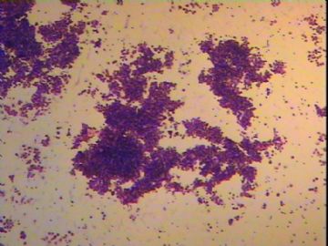
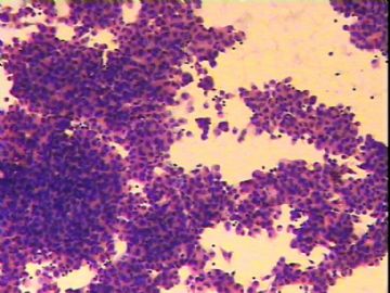
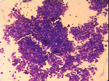
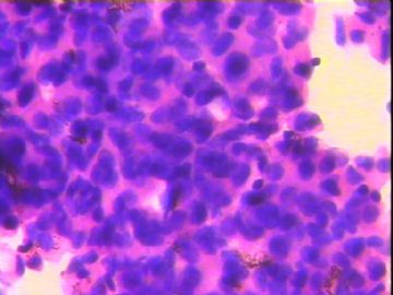
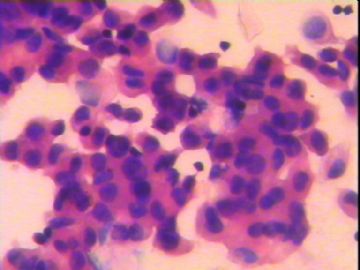
- 胸水是癌细胞吗?还是增生的间皮细胞?
-
The cytologic details of the photos are not very high. While carcinoma and reactive mesothelial hyperplasia are the two main differential diagnoses, cytologic details are essential in their distinction. In addition, the hypercellularity and complex papillary groups of the cells present also suggest malignant mesothelioma. If there is enough specimen to prepare a cell block, immunohistochemical stains are very helpful. If not, chect CT scans need to be examined carefully for any pleural irregularity or tumors. If there is abnormality, a thoracic surgeon has to be consulted to do a thoracoscopic biopsy of any suspicious tumors on pleural surface.

聞道有先後,術業有專攻

