


| 图片: | |
|---|---|
| 名称: | |
| 描述: | |
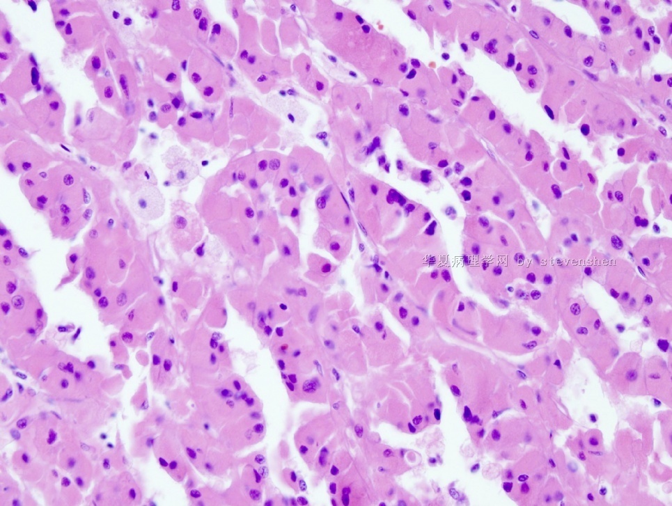
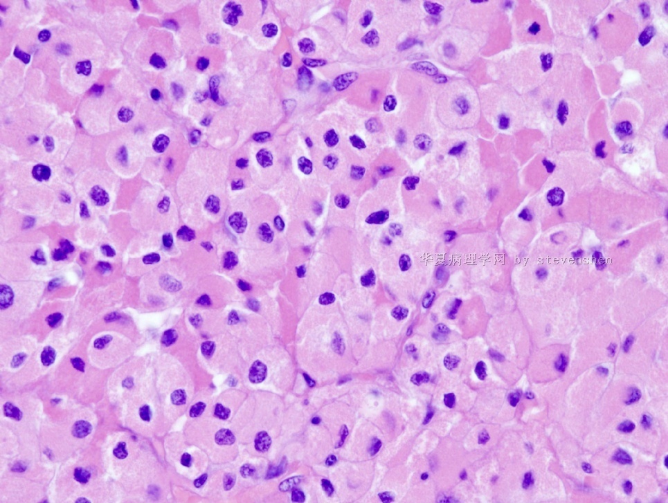
- 70 year old man with 4.0 cm renal mass
-
stevenshen 离线
- 帖子:343
- 粉蓝豆:2
- 经验:343
- 注册时间:2008-06-03
- 加关注 | 发消息
 图1
图1 图2
图2 图3
图3 图4
图4 图5
图5
| 姓 名: | ××× | 性别: | Male | 年龄: | 70 |
| 标本名称: | nephrectomy | ||||
| 简要病史: | renal mass | ||||
| 肉眼检查: | well-circumscribed homogenous brown mass with focal hemorrhage | ||||
Your frozen diagnosis and best guess for final diagnosis and why?
标签:
×参考诊断
嫌色细胞癌(Chromophobe renal cell carcinoma)
-
wangdingding 离线
- 帖子:1474
- 粉蓝豆:98
- 经验:6042
- 注册时间:2006-10-19
- 加关注 | 发消息
-
What about oncocytoma? The homogeneous brown color of the tumor, gross and microscopic circumscription, plump eosinophilic cytoplasm and relatively small nuclei are all consistent with it. Certainly one should consider the granular cell variant of clear cell renal cell carcinoma and chromophobe renal cell carcinoma. In the former, features of clear cell carcinoma are usually found focally under the microscope and its gross appearance is usually heterogeneous yellow to tan to brown. As for the latter, the cytoplasm is never granular (more likely "clear" on FS) and intercellular borders are very distinct (not seen in this case).

聞道有先後,術業有專攻
-
pathseeker 离线

- 帖子:20
- 粉蓝豆:1
- 经验:20
- 注册时间:2009-11-17
- 加关注 | 发消息
-
stevenshen 离线
- 帖子:343
- 粉蓝豆:2
- 经验:343
- 注册时间:2008-06-03
- 加关注 | 发消息
-
The permanent section shows features that are not consistent with a pure oncocytoma. The differential diagnoses go back to granular cell variant of clear cell renal cell carcinoma and eosinophilic variant of chromophobe renal cell carcinoma. The distinction requires careful microscopic examination, which usually would suffice to tell the two apart. Without such possibility here and relying on selected photomicrographs, investigative staining is needed - including CD10, CD117, vimentin, CK7 and RCC-Ma immunostains and Hale's colloidal iron stain. I personally favor this is a case of granular cell variant of clear cell RCC.
聞道有先後,術業有專攻
-
liguoxia71 离线

- 帖子:4174
- 粉蓝豆:3122
- 经验:4677
- 注册时间:2007-04-01
- 加关注 | 发消息
-
stevenshen 离线
- 帖子:343
- 粉蓝豆:2
- 经验:343
- 注册时间:2008-06-03
- 加关注 | 发消息
- Thanks for all the abover discussion. I don't have much to add.
- Intraoperative frozen section diagnosis is usually not necessary for renal tumor surgery.
- In some situations such as this case, a definitive diagnosis may be difficult.
- The main differential diagnoses for this case are: oncocytoma vs. chromophobe RCC (eosinophilic variant) vs clear cell RCC (with granular cells).
- Frozen diagnosis: oncocytic renal neoplasm, chromophobe versus oncocytoma
- Final diagnosis: chromophobe RCC, eosinophilic variant
-
分享知识点:Chromophobe renal cell carcinoma
- Large polygonal cells with pale, flocculent cytoplasm and very distinct plasmalemmal outlines. The cells are arranged in solid nests or tubules. Ultrastructurally, the flocculence of the cytoplasm has been found to be due to large numbers of minute vesicles. Hale's colloid iron histochemical characteristically colors the cytoplasm diffusely blue. Although two histological variants are recognized, a typical and an eosinophilic one (increased numbers of mitochondria), both cell types may occur in the same tumor.
- Rare example of composite oncocytoma/chromophobe RCC tumors have been described leading, to the hypothesis that the two types of tumors may be related. Recently, immunohistochemical staining for the RCC antigen and CD10 has been proposed as a possiblly useful adjunct in the differential diagnoses of chromophobe RCC vs clear cell type RCC and chromophobe RCC vs oncocytoma. There is evidence that chromophobe RCCs are less likely to behave aggressively than otherwise similar clear cell type RCCs.
-
本帖最后由 于 2010-02-21 20:22:00 编辑
本例嫌色细胞癌的嗜酸细胞亚型的诊断完全成立。诊断时,是需要与嗜酸细胞腺瘤鉴别。
1.临床表现和影像学一般很难鉴别,但在影像学上嗜酸细胞腺瘤形状相对规则,可呈轮辐状。
2.大体观:大体标本也有可能增加诊断的信心,嫌色细胞癌的嗜酸细胞亚型的色彩可表现不均一。而嗜酸细胞腺瘤大体观就如同肝脏局灶结节状增生一样,肿块常生长在靠近器官的被摸下并向表面凸出,可出现中央放射状星芒状疤痕(灰白的结缔组织间质区)。瘤体切面呈一致的棕褐色。
3.组织病理:组织学是一个基本的判断。嫌色细胞癌嗜酸细胞亚型或多或少可见到嫌色细胞(表现为胞浆淡染或絮网状或在嗜酸细胞腺中见到核周晕,这是由于胞浆内含有大量的小的液泡所致。而嗜酸细胞腺瘤的胞浆嗜酸性相对比较一致(胞浆颗粒强嗜酸性),而且巢状结构(或腺囊状)的边缘比较规则,多数还有疏松水肿的间质背景。细胞核的形态对鉴别诊断也很重要,嗜酸性腺瘤的胞核较深染,核仁不明显,虽偶可出现怪异大核,但一般不应出现核分裂、无乳头状结构和成片透明细胞或梭形细胞,无明显的血管浸润等(广泛取材对于鉴别很重要)。
4.组织化学:Hale胶体铁染色是个很好的鉴别诊断方法,嫌色细胞癌染色阳性(胞浆呈蓝色),嗜酸细胞腺瘤染色阴性。
.5.IHC:IHC标记的鉴别意义不大。但RCC一般可表达Vimentin,而嗜酸细胞腺瘤一般不表达Vimentin。
仅供参考!
- 王军臣


