| 图片: | |
|---|---|
| 名称: | |
| 描述: | |
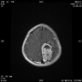
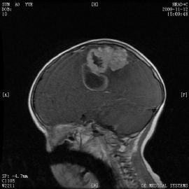
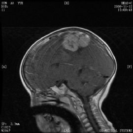
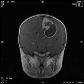
- 小孩左顶叶肿瘤
-
liangjinjun 离线
- 帖子:2328
- 粉蓝豆:2
- 经验:2457
- 注册时间:2007-08-07
- 加关注 | 发消息
| 姓 名: | ××× | 性别: | 女 | 年龄: | 4岁 |
| 标本名称: | 左侧顶叶肿块 | ||||
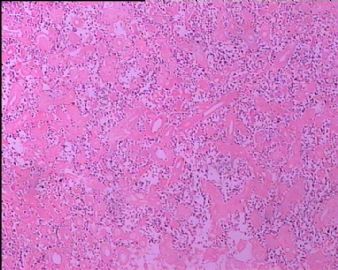
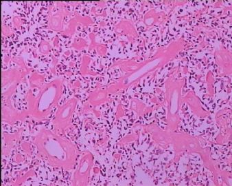
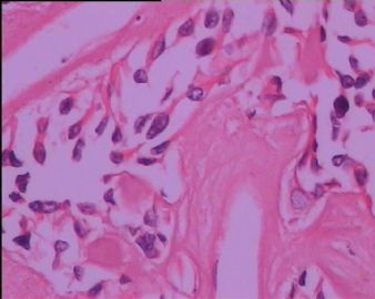
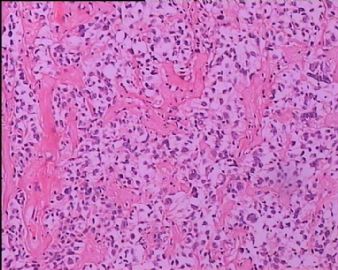
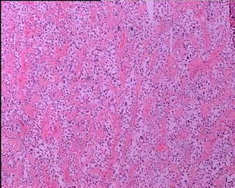
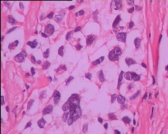
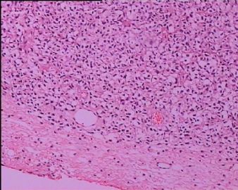
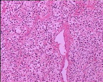
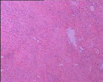
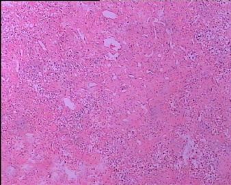
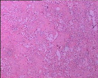
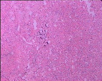
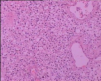
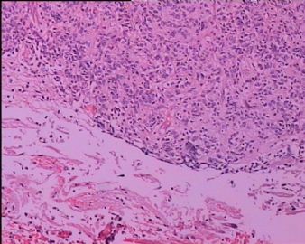
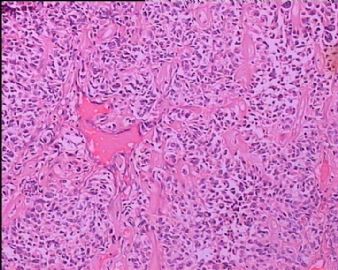
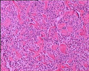
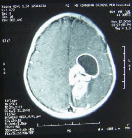
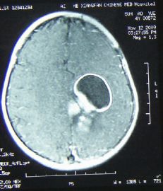
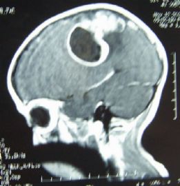
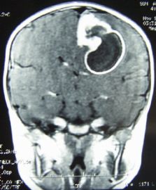
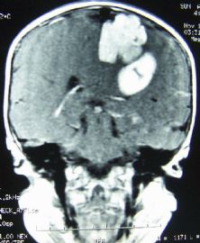
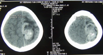
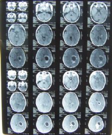
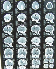
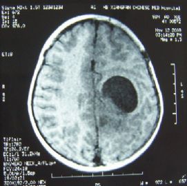
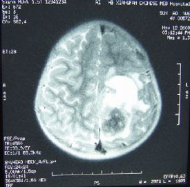
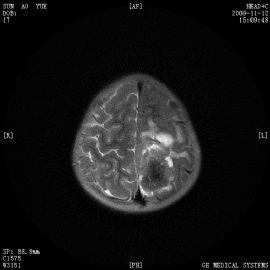
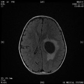
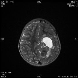
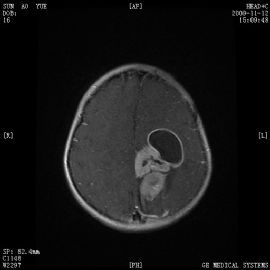
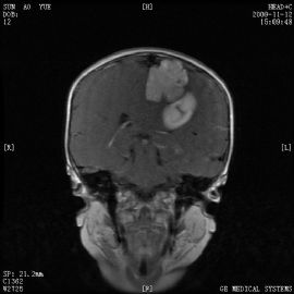
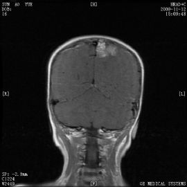
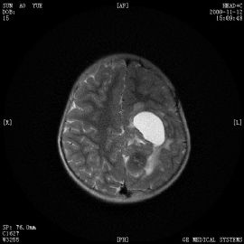
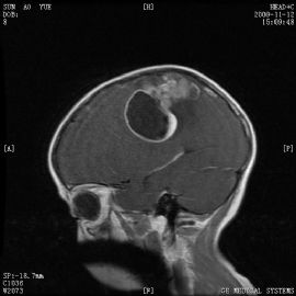
| 简要病史: | 左侧顶叶肿块,一侧与脑膜相连,伴囊性变。 | ||||
| 肉眼检查: | 送检4X4X3CM一堆,灰白灰红色,质脆。 | ||||
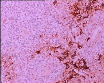
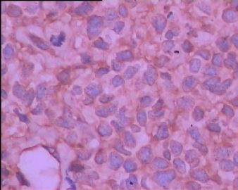
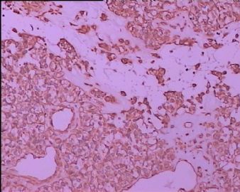
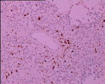
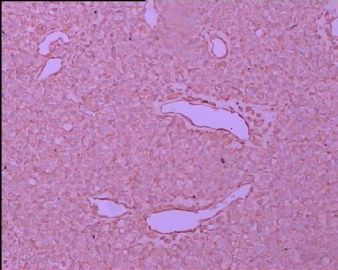
免疫组化:EMA局灶+、GFAP可疑+、VIM+、KI67+15%
(免疫组化图依次为EMA、GFAP高倍、GFAP中倍、KI67、VIM)
标签:
-
本帖最后由 于 2009-11-23 19:58:00 编辑

- 梁晋军
×参考诊断
-
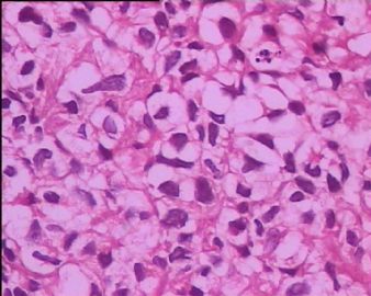
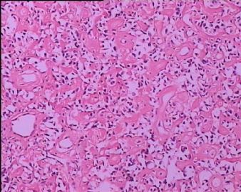
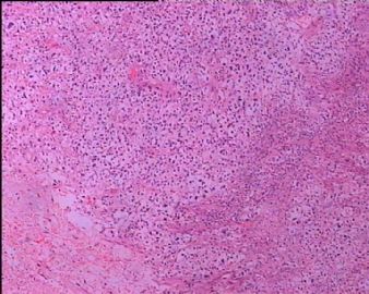
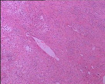
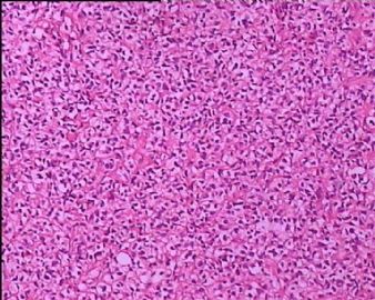
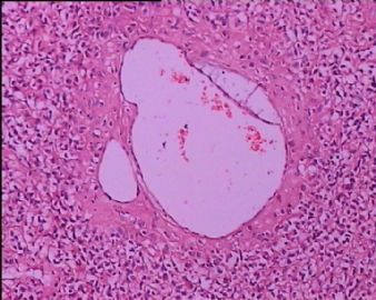
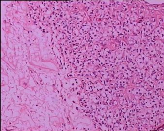
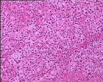
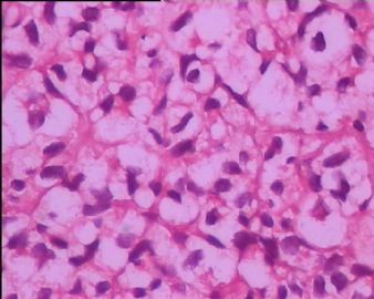
This is a difficult case to interpret. From the photos uploaded, I can tell what this malignant neoplasm is not, but I cannot tell what it is for sure. Many cells have clear or vacuolated cytoplasm, and there are many thick collagenous fibers and sinusoidal vessels between cells. Cells in Figures 18-20 appear somewhat different from the rest. The patient's young age does not present many differential diagnoses, but I can not imagine this being a benign meningioma, hemangioblastoma, pilocytic astrocytoma, desmoplastic infantile ganglioglioma, desmoplactic cerebral astrocytoma of infancy, other types of ganglioglioma, oligodendroglioma, ependymoma or fibrillary astrocytoma. It does suggest hemangiopericytoma. I would need more information (pre-surgical MRI) before I can give an opinion.

聞道有先後,術業有專攻
-
liangjinjun 离线
- 帖子:2328
- 粉蓝豆:2
- 经验:2457
- 注册时间:2007-08-07
- 加关注 | 发消息
-
liangjinjun 离线
- 帖子:2328
- 粉蓝豆:2
- 经验:2457
- 注册时间:2007-08-07
- 加关注 | 发消息
-
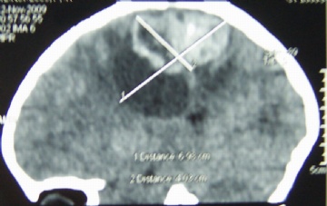
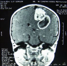
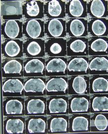
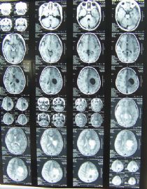
Thank you for uploading the neuroimages expeditiously. From them, I see an intra-axial, fairly well cirecumscribed and lobulated, cystic/solid parenchymal tumor without much surrounding edema. This remains a difficult malignant neoplasm to classify and grade. In addition to showing focal cautery artefacts, the photomicrographs show thick and broad collageous fibers and large gaping sinusoidal vessels between neoplastic cells. Large areas of fibrosis and focal calcification are seen. I wonder if more photos of well preserved areas stained by HE would help or not (especially near where photos 18~20 were taken that show cells with more pleomorphic nuclei and no vacuolated cytoplasm). GFAP immunoreactivity seems real and diffuse, but EMA immunoreactivity is questionable. I can safely conclude that this is not a clear cell meningioma, hemangioblastoma, desmoplastic cerebral astrocytoma of infancy, desmoplastic infantile ganglioglioma, and pleomorphic xanthoastrocytoma. Hemangiopericytoma may show clear cells, but it is almost never cystic. Pilocytic astrocytoma remains an important neoplasm to rule out. It can contain many clear cells like in this case, so we have to look for eosinophilic granular bodies, Rosenthal fibers and biphasic growth pattern in any area. An oligodendroglioma is possible, but the blood vessel pattern seen here is not consistent with delicate capillaries seen in oligodendroglioma. This may also be a rare form of glioneuronal tumor that cannot be classified easily. Finally, astroblastoma should be considered in such a young child. It usually would show striking papillary growth in addition to hyalinized fibrosis. So, the bottom line is with the images available so far I DON'T KNOW WHAT THIS IS. It would help to examine all the slides under the microscope to appreciate histopathology first hand. If you have no additional good diagnostic idea, you should consider sending it out for an expert second opinion. Alternatively, scan a few representative HE slides into digital files for telepathology consultation. Hope my discussion helps.

聞道有先後,術業有專攻
-
liangjinjun 离线
- 帖子:2328
- 粉蓝豆:2
- 经验:2457
- 注册时间:2007-08-07
- 加关注 | 发消息

