| 图片: | |
|---|---|
| 名称: | |
| 描述: | |
- 右额顶叶占位
-
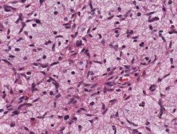
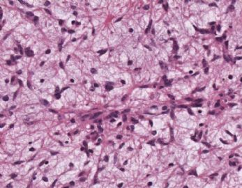
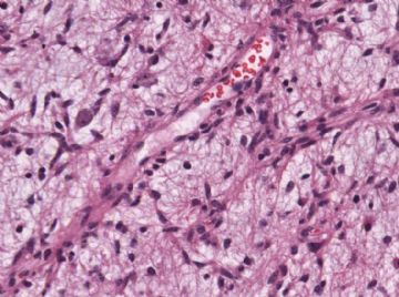
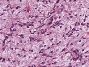
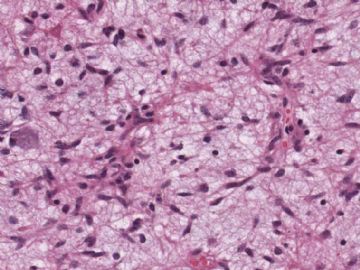
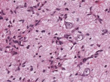
The photos show an astrocytoma, with scattered large neurons seen focally to suggest ganglioglioma. Differential diagnoses include WHO grade I pilocytic astrocytoma, WHO grade II pilomyxoid astrocytoma, WHO grade II (or, less likely, WHO grade III) diffuse infiltrating (fibrillary) astrocytoma, and WHO grade II (or, less likely, WHO grade III) ganglioglioma. To make the distinction between all these, one needs to observe for (1) eosinophilic granular bodies, (2) biphasic growth pattern, (3) microcysts, (4) Rosenthal fibers, (5) interface between tumor and surrounding parenchyma (demarcated or infiltrative), (6) mitotic counts, (7) any necrosis, and (8) vascular/endothelial proliferation. Based on these few photos alone it is insufficient to make a final assessment.

聞道有先後,術業有專攻

