| 图片: | |
|---|---|
| 名称: | |
| 描述: | |
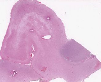
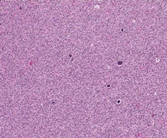
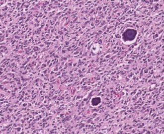
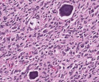
- 左额叶占位
-
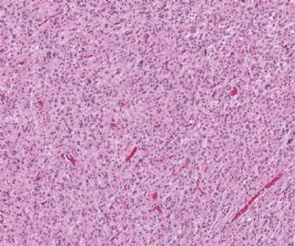
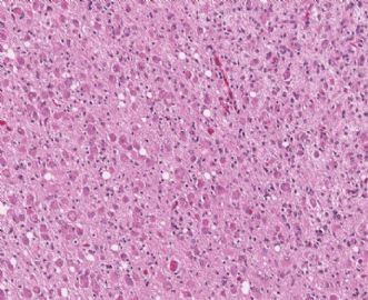
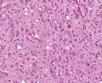
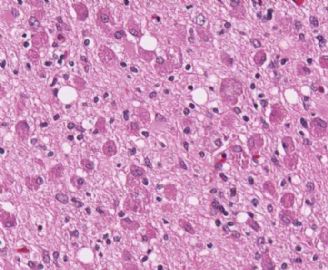
Although mitotic figures are difficult to identify on few photos, the cellularity and cytology of this case are that of WHO grade III anaplastic astrocytoma or glioblastoma, depending whether vascular/endothelial proliferation is observed focally or not. I do not seen classic tumor necrosis. The neoplastic cells with eosinophilic cytoplasmic granules are seen in some cases of gliomas (astrocytomas or oligodendrogliomas) of various grades, and have been considered as a degenerative change. They should not be confused with eosinophilic granular bodies.

聞道有先後,術業有專攻

