| 图片: | |
|---|---|
| 名称: | |
| 描述: | |
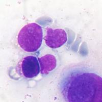
- 胸水,请各位老师会诊是否是癌细胞,谢谢!
-
junzi003981 离线
- 帖子:1469
- 粉蓝豆:234
- 经验:3249
- 注册时间:2008-09-07
- 加关注 | 发消息
这例涂片首先是干固定的,细胞有退变,看不清核染色质、核仁和核膜,这对诊断带来很大的困难。
从细胞核浆比和核形上看,大多数细胞以间皮增生可能性大,但那些退变的核偏位的细胞,胞浆好象还有粘液,我不放心。所以这一例我只能报找到少量可疑癌细胞或核异质细胞。
建议:
1、希望能湿固定制片,你的涂片质量很好,这样的片子涂好后直接放入95%酒精肯定没问题,如果不放心,可以多做几张,二张干固定,二张湿固定,比较一下。
2、可以做AB染色,这是最方便和有效的方法。
3、免疫组化和细胞蜡块要看你的条件了。

- 本人观点纯属个人偏见,希望大家独立判断,正确引用!中华病理技术网:http://www.zhbljs.com/
-
本帖最后由 于 2009-10-23 14:23:00 编辑
| 以下是引用cqzhao在2009-10-23 11:15:00的发言: It is strange that all cells are single or isolated. |
Dr. Zhao has found the key feature of the cytology. If you want to make a definite diagnosis of adenocarcinoma, the 3-D particles of cells ARE VERY IMPORTANT. For the mesothelial cell might mimic the morphology of tumors as you want.
To my knowledge, I can not call it a tumor or metastasis.

- 用心做事、真情做人!
| 以下是引用海上明月在2009-10-22 17:42:00的发言:
见高度核异质细胞,疑癌。 年老患者单侧血性胸水是凶多吉少。可是,细胞相对比较弥散,很难见到粘着成团成蔟的、乳头状的或腺样的结构。甚至许多细胞可能是增生的间皮细胞。需要再送胸水复查,必要时制作细胞块做IHC标记鉴别诊断。 |
Agree above. Do two stains, BerEp4 (for epithelial cells) and calretinin (间皮细胞) if you can. Mostly you can get the answer. If both are negative, do CD68 (histiocytes) and LCA (WBC, lymphocytes).
I cannot make definite diagnosis if IHC cannot be performed.
-
skyliutong 离线
- 帖子:497
- 粉蓝豆:189
- 经验:878
- 注册时间:2009-02-14
- 加关注 | 发消息


