| 图片: | |
|---|---|
| 名称: | |
| 描述: | |
- 72 year old with a large thigh mass
-
stevenshen 离线
- 帖子:343
- 粉蓝豆:2
- 经验:343
- 注册时间:2008-06-03
- 加关注 | 发消息
| 姓 名: | ××× | 性别: | 72 | 年龄: | male |
| 标本名称: | Thigh mass (9 cm) | ||||
| 简要病史: | |||||
| 肉眼检查: | Homogeneous fleshy mass with focal cystic change | ||||
Frozen section interpretation and diagnosis?
Have difficulties to upload the photomicrograph..will try another time...
标签:
-
本帖最后由 于 2009-10-31 06:19:00 编辑
×参考诊断
Solitary fibrous tumor
-
stevenshen 离线
- 帖子:343
- 粉蓝豆:2
- 经验:343
- 注册时间:2008-06-03
- 加关注 | 发消息
- Although there are not many responses, but all the discussion are to the point.
- For this particular case, I think 冰冻切片报告间叶源性肿瘤 will be sufficient, unless one can absolutely sure about the diagnosis. It will be dangerous to make a incorrect diagnosis that lead to unneccesary radical surgery. One has to be cautious to make a definite "benign" or malignant diagnosis.
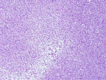
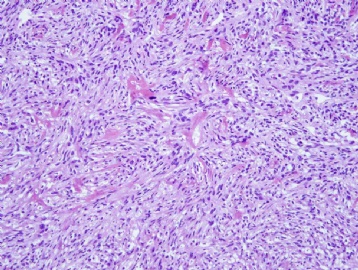
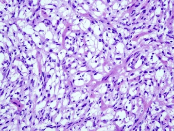
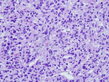
- Frozen diagnosis: cellular spindle cell neoplasm
- Final diagnosis: solitary fibrous tumor (CD34 diffusely and strongly positive)
- Thanks to Dr. Ma for the excellent comments!
-
My impression based on the FS photomicrographs was made boldly with a hunch and aided by the excellent FS quality. In real practice I would have been more cautious. The high cellularity of the neoplasm, a vague storiform growth pattern, vacuolated cytoplasm seen at least focally, thick collagenous fibers, and many collapsed small vessels (capillaries). While all of these are consistent with a few types of soft tissue neoplasm, I was most impressed by the last high power photo of FS - it looks just like the hemangiopericytoma I am familiar with. The anatomic location and patient's old age should bring out a few differential diagnoses with such a hypercellular spindle cell neoplasm. Others aside, I will say that cells in sarcomatoid synovial sarcoma and malignant peripheral nerve sheath tumor usually are more uniform in the nuclear size and shape (elongated or oval) and more intersecting and fascicular in architecture. Cells in hemangiopericytomas have moderately pleomorphic nuclear size and shape, and they can display storiform growth and clear cells focally. Immunohistochemical confirmation on permanent sections is needed without exception.

聞道有先後,術業有專攻
-
stevenshen 离线
- 帖子:343
- 粉蓝豆:2
- 经验:343
- 注册时间:2008-06-03
- 加关注 | 发消息
-
pathseeker 离线
- 帖子:20
- 粉蓝豆:1
- 经验:20
- 注册时间:2009-11-17
- 加关注 | 发消息
-
stevenshen 离线
- 帖子:343
- 粉蓝豆:2
- 经验:343
- 注册时间:2008-06-03
- 加关注 | 发消息


