| 图片: | |
|---|---|
| 名称: | |
| 描述: | |
- 右额叶肿块
-
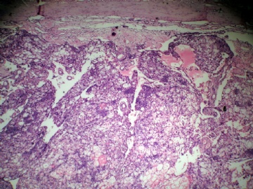
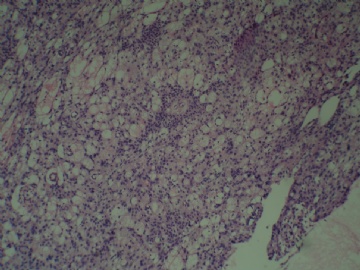
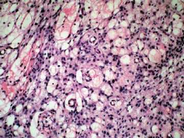
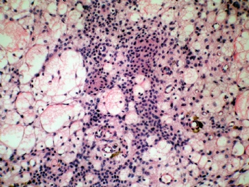
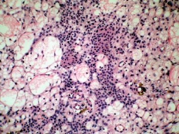
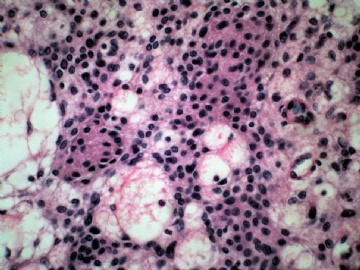
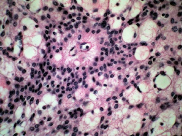
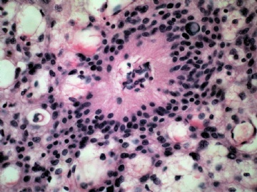
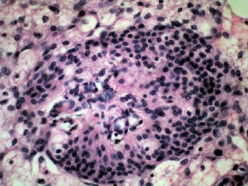
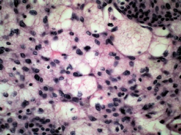
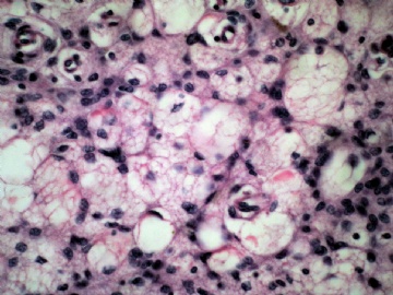
This is a case of microcystic/lipidized meningioma, WHO grade I. The demarcation from brain tissue is sharp (photo 1) with focal calcification (photo 1), vague whorl formation (photos 5 and 6), and uniform cells without atypical features. Perivascular pseudorosettes (photos 7 and 8) are not diagnostic of ependymoma. They can be seen in various gliomas (astrocytomas and ependymomas) and meningiomas. If unsure, do PR and EMA immunostains to confirm it.

聞道有先後,術業有專攻

