共1页/6条首页上一页1下一页尾页
回复:13 阅读:2449
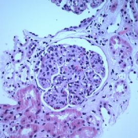
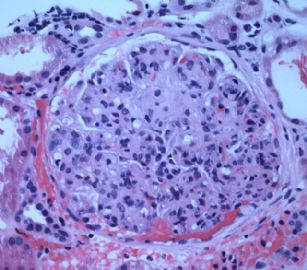
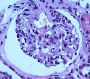
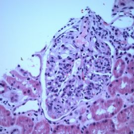
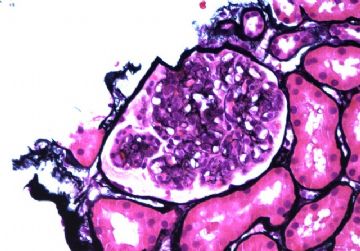
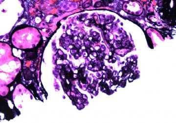
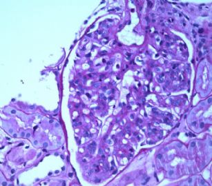
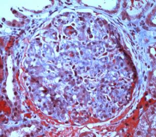
Adult with proteinuria.

quhong 离线

帖子:493
粉蓝豆:24
经验:953
注册时间:2009-02-22
加关注  |  发消息
楼主 发表于 2009-09-07 01:28|举报|关注(0)
浏览排序[ 顺序 逆序 楼主 支持 精彩 ]  快捷回复
姓    名: ××× 性别:   年龄:  adult
标本名称:  
简要病史:  Proteinuria (3.0 grams).
肉眼检查:  

Immunofluorescence microscopy is completely negative. This is unexpected.


I am waiting for electron microscopy results.

  • Adult with proteinuria.图1
    图1
  • Adult with proteinuria.图2
    图2
  • Adult with proteinuria.图3
    图3
  • Adult with proteinuria.图4
    图4
  • Adult with proteinuria.图5
    图5
  • Adult with proteinuria.图6
    图6
  • Adult with proteinuria.图7
    图7
  • Adult with proteinuria.图8
    图8
标签:
0
添加参考诊断
×参考诊断
  

quhong 离线

帖子:493
粉蓝豆:24
经验:953
注册时间:2009-02-22
加关注  |  发消息
1 楼    发表于2009-09-09 21:38:00举报|引用
返回顶部 | 快捷回复
本帖最后由 于 2009-09-09 21:43:00 编辑
以下是引用geng72在2009-9-9 16:22:00的发言:

 此例患者突出的病理改变是:毛细血管内细胞的增生伴中性粒细胞的浸润,还可见到少量嗜酸性粒细胞,毛细血管襻呈分叶状改变,局部可见血管襻的纤维素样坏死,肾间质有炎症细胞浸润和血管壁的纤维素样坏死。需要考虑的诊断有:

1、毛细血管内增生性肾小球肾炎,支持点:组织学改变

    不支持点:临床主要以大量蛋白尿为主,免疫荧光阴性

    但对于约5%的毛细血管内增生性肾小球肾炎患者会以大量蛋白尿为主要表现,而一些可能为病毒感染引起的毛细血管内增生性肾小球肾炎可以荧光阴性,电镜无驼峰样改变,

2、一些IgA肾病及继发性肾小球肾炎,如系统性红斑狼疮,冷凝球蛋白血症,等,这些可以通过临床及免疫荧光排除。

3、血栓性微血管病,我们曾遇到几例由于妊高症引起的血栓性微血管病,可以表现为内皮细胞的增生、肿胀,但一般无明显炎症细胞浸润,基底膜会水肿、增厚,电镜检查会有帮助,一般血栓性微血管病还会有更多的临床表现

总之,这是一个很特殊的病例,我还是主要考虑因感染引起的毛细血管内增生性肾小球肾炎,不一定是链球菌感染,可能是其它的病原体感染引起的,期待最终的结果及电镜检查。

Dr. Geng: I think your analysis is right. This is not a thrombotic microangiopathy. The below is the EM. Let us know your thought.

名称:图1
描述:图1

名称:图2
描述:图2

名称:图3
描述:图3

名称:图4
描述:图4

名称:图5
描述:图5

名称:图6
描述:图6

名称:图7
描述:图7

名称:图8
描述:图8

名称:图9
描述:图9

名称:图10
描述:图10

名称:图11
描述:图11

名称:图12
描述:图12

名称:图13
描述:图13

名称:图14
描述:图14

名称:图15
描述:图15
0
回复

quhong 离线

帖子:493
粉蓝豆:24
经验:953
注册时间:2009-02-22
加关注  |  发消息
2 楼    发表于2009-09-09 21:36:00举报|引用
返回顶部 | 快捷回复
本帖最后由 于 2009-09-09 21:44:00 编辑
以下是引用wfbjwt在2009-9-8 22:00:00的发言:

 血管炎,多结节性 OR 坏死性 OR others?

Morphologically 血管炎 is there. At this moment, I am not sure it is polyarteritis nodosa. Patient's serology for ANCA is negative. Yesterday I read a clinical consultation report, which states that this patient is "positive for Wagner disease, hypertension and diabetes". but I have not seen a single crescent in this biopsy (total 29 glomeruli).I just got the EM, which shows endocapillary proliferation. Initially I thought this was a chronic form of thrombotic microangiopathy. The EM does not support it.
0
回复

quhong 离线

帖子:493
粉蓝豆:24
经验:953
注册时间:2009-02-22
加关注  |  发消息
3 楼    发表于2009-09-08 21:29:00举报|引用
返回顶部 | 快捷回复
本帖最后由 于 2009-09-08 21:33:00 编辑  After noticing the vascular changes, I realized that the glomerular changes actually were there. I simply did not see them(!).

名称:图1
描述:图1

名称:图2
描述:图2

名称:图3
描述:图3
0
回复

quhong 离线

帖子:493
粉蓝豆:24
经验:953
注册时间:2009-02-22
加关注  |  发消息
4 楼    发表于2009-09-08 21:20:00举报|引用
返回顶部 | 快捷回复
本帖最后由 于 2009-09-08 21:23:00 编辑 The past weekend I took this case home and carefully reviewed the slides again. Now I have diagnostic findings. The following pictures are most important ones:

名称:图1
描述:图1

名称:图2
描述:图2

名称:图3
描述:图3

名称:图4
描述:图4

名称:图5
描述:图5
0
回复

quhong 离线

帖子:493
粉蓝豆:24
经验:953
注册时间:2009-02-22
加关注  |  发消息
5 楼    发表于2009-09-07 21:08:00举报|引用
返回顶部 | 快捷回复
以下是引用wfbjwt在2009-9-7 18:17:00的发言:

 类似分叶状肾小球肾炎的形态。


I have the same feeling. Now I count on EM to give me some helpful findings.

0
回复

quhong 离线

帖子:493
粉蓝豆:24
经验:953
注册时间:2009-02-22
加关注  |  发消息
6 楼    发表于2009-09-07 21:05:00举报|引用
返回顶部 | 快捷回复
以下是引用tata在2009-9-7 17:43:00的发言:

 镜下表现为毛细血管内增生性肾小球肾炎改变,中性粒细胞浸润明显,小球呈分页状外观。如果是链球菌感染引起,一般IF染色应该有IgG、C3等免疫复合物沉积。本例以大量蛋白尿为唯一表现吗?等待更多的资料以确定可能的诊断!


I have talked to the nenphrologist twice. Proteinuria is the only indication for this patient's biopsy. I am also puzzled by the negative immunofluorescence stains!  Thank you for sharing your thought with us.

0
回复
回复:13 阅读:2449
共1页/6条首页上一页1下一页尾页
【免责声明】讨论内容仅作学术交流之用,不作为诊疗依据,由此而引起的法律问题作者及本站不承担任何责任。
快速回复
进入高级回复
您最多可输入10000个汉字,按 "Ctrl" + "Enter" 直接发送
搜索回复/乘电梯 ×
按内容
按会员
乘电梯
合作伙伴
友情链接