| 图片: | |
|---|---|
| 名称: | |
| 描述: | |
- B2242左乳肿物 (29楼新传免疫组化,有当没有看看吧)
| 姓 名: | ××× | 性别: | 女 | 年龄: | 55 |
| 标本名称: | |||||
| 简要病史: | |||||
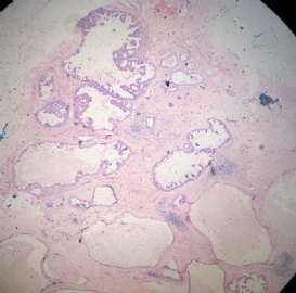
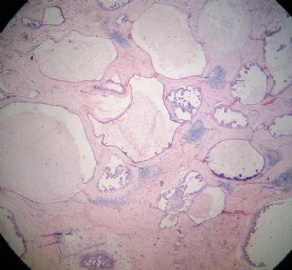
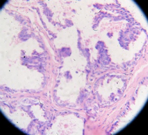
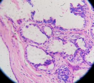
| 肉眼检查: | 灰白灰黄组织一块,3.8*2.7*2.4厘米,切面灰白灰黄,质硬,有乳汁样物流出。 | ||||
-
本帖最后由 于 2009-09-28 20:47:00 编辑
相关帖子
- • 乳腺包块
- • 左乳癌标本乳头一个导管内的病变
- • 乳腺两个相邻导管内的病变
- • 乳腺肿物
- • 乳腺肿物,请各位老师帮忙会诊
- • 女 46岁发现左乳腺肿块一月余
- • 乳腺包块。33岁
- • 左乳肿块,协助诊断
- • 乳腺肿物
- • 乳腺肿物
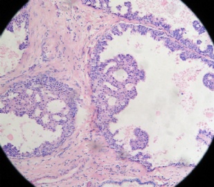
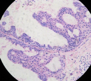
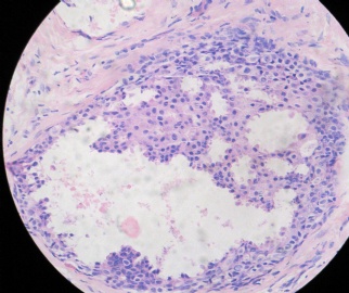
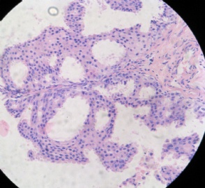
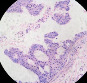
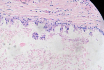
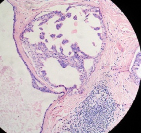
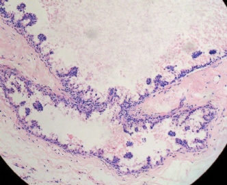
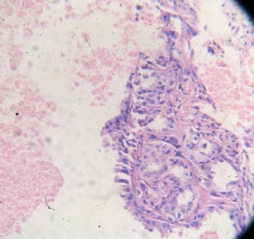
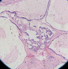
From low power we can appreciate focal proliferation in FCC-like background (it can be FA if it is demarcated mass). The photos from high power demonstrate micropapillary growth patterns with relative uniform cells. I favor a diagnosis of atypical ductal hyperplasia. I feel it is not enough for DCIS.
Just for your reference.
-
liangjinjun 离线
- 帖子:2328
- 粉蓝豆:2
- 经验:2457
- 注册时间:2007-08-07
- 加关注 | 发消息
| 以下是引用cqzhao在2009-9-8 2:29:00的发言:
From low power we can appreciate focal proliferation in FCC-like background (it can be FA if it is demarcated mass). The photos from high power demonstrate micropapillary growth patterns with relative uniform cells. I favor a diagnosis of atypical ductal hyperplasia. I feel it is not enough for DCIS. Just for your reference. |
-
taotaochan 离线
- 帖子:131
- 粉蓝豆:22
- 经验:131
- 注册时间:2007-03-17
- 加关注 | 发消息
| 以下是引用绝世好片在2009-9-9 22:17:00的发言:
|
We have so many 版主 and 助理 here or others with good english. No person can help?
thanks, cz
| 以下是引用cqzhao在2009-9-12 19:10:00的发言:
We have so many 版主 and 助理 here or others with good english. No person can help? thanks, cz |
-
From low power we can appreciate focal proliferation in FCC-like background (it can be FA if it is demarcated mass). The photos from high power demonstrate micropapillary growth patterns with relative uniform cells. I favor a diagnosis of atypical ductal hyperplasia. I feel it is not enough for DCIS.
Just for your reference
从低倍镜下我们能看出在纤维囊性腺病背景中有局灶性增生(如果肿块境界清楚则可能是纤维腺瘤)。高倍镜下病变可见微乳头状生长,细胞大小相对一致,我考虑非典型导管增生,不够DCIS。
仅供参考。
| 以下是引用微尔笑在2009-9-16 12:04:00的发言:
|
Thank Dr. 微尔笑.
Hope our pathologists can still spend some time on pathology.

