
 - metastatic alveolar soft part sarcoma图1")
 - metastatic alveolar soft part sarcoma图2")
 - metastatic alveolar soft part sarcoma图3")
 - metastatic alveolar soft part sarcoma图4")
 - metastatic alveolar soft part sarcoma图5")
 - metastatic alveolar soft part sarcoma图6")
 - metastatic alveolar soft part sarcoma图7")
 - metastatic alveolar soft part sarcoma图8")


| 图片: | |
|---|---|
| 名称: | |
| 描述: | |
- NP (8) - metastatic alveolar soft part sarcoma
-
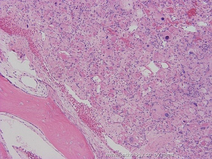
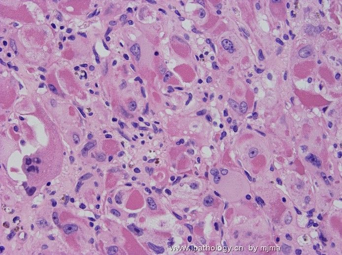
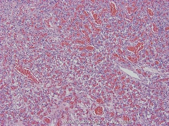
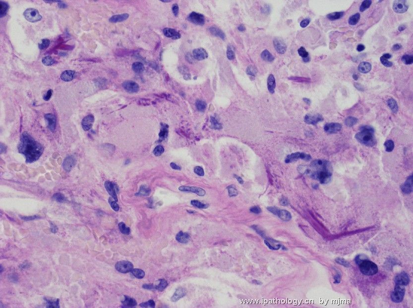
A 25-year-old man presented with new seizure disorder, and was found to have multiple enhancing lesions in the brain, spinal cord, none (vertebral column) and liver. The photos are taken from a resected left temporal lobe brain tumor. Figures 1~3 are from frozen section (HE) at magnification 10x, 20x and 40x. Figures 4~8 are from HE-stained paraffin section at magnification 4x, 10x, 20x, 40x and 60x. What are your differential diagnoses? What further workup do you suggest?
 - metastatic alveolar soft part sarcoma图1") 图1
图1 - metastatic alveolar soft part sarcoma图2") 图2
图2 - metastatic alveolar soft part sarcoma图3") 图3
图3 - metastatic alveolar soft part sarcoma图4") 图4
图4 - metastatic alveolar soft part sarcoma图5") 图5
图5 - metastatic alveolar soft part sarcoma图6") 图6
图6 - metastatic alveolar soft part sarcoma图7") 图7
图7 - metastatic alveolar soft part sarcoma图8") 图8
图8
标签:
-
本帖最后由 于 2008-02-17 22:31:00 编辑

聞道有先後,術業有專攻
×参考诊断
-
本帖最后由 于 2007-05-24 20:47:00 编辑
A 25-year-old man presented with new seizure disorder, and was found to have multiple enhancing lesions in the brain, spinal cord, none (vertebral column) and liver. 患者、男、25岁,癫痫发作,作CT增强发现脑、脊髓、脊柱和肝脏有多个病灶。The photos are taken from a resected left temporal lobe brain tumor. 照片取自左颞叶脑肿瘤处活检。Figures 1~3 are from frozen section (HE) at magnification 10x, 20x and 40x. 图1-3是冰冻切片、HE染色,放大100、200、400倍。Figures 4~8 are from HE-stained paraffin section at magnification 4x, 10x, 20x, 40x and 60x.图4-8是常规石腊切片,HE染色,放大40、100、200、400、600倍。 What are your differential diagnoses? 看您需要做那些鉴别诊断?What further workup do you suggest? 您提议再做些什么?
-
wangdingding 离线
- 帖子:1474
- 粉蓝豆:98
- 经验:6042
- 注册时间:2006-10-19
- 加关注 | 发消息
-
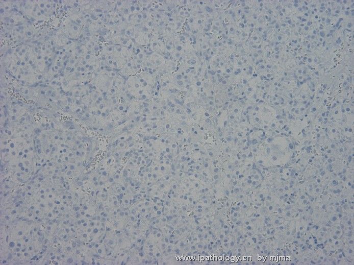
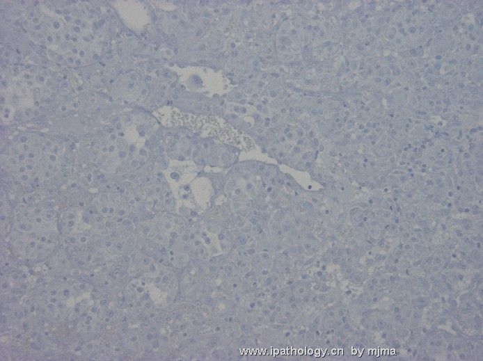
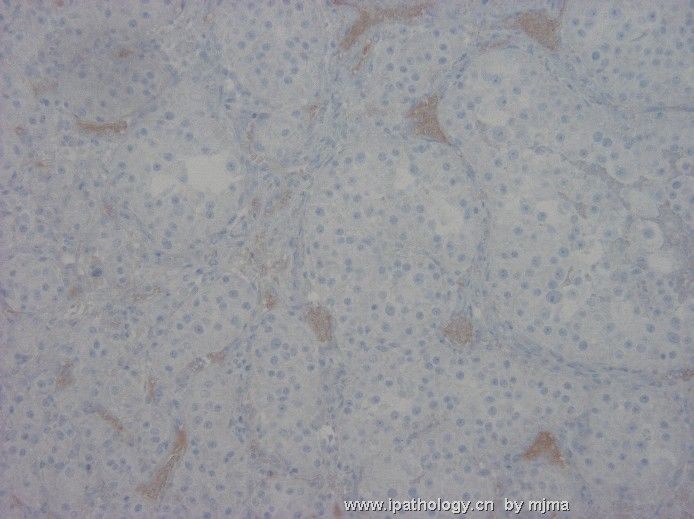
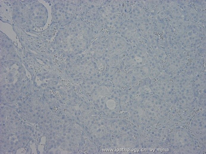
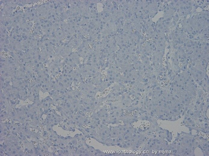
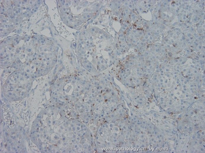
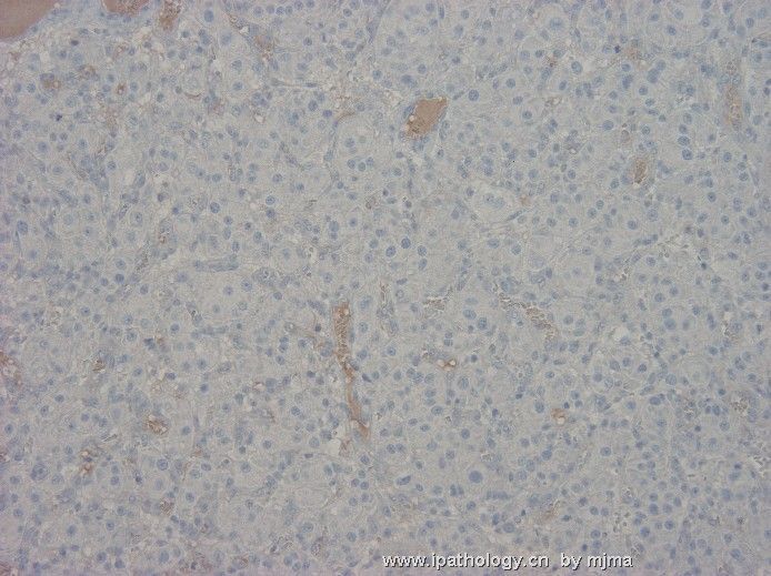
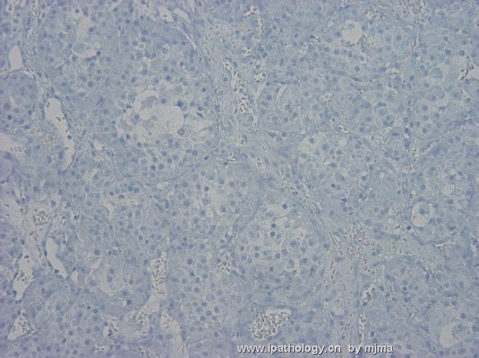
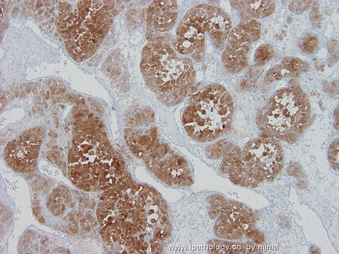
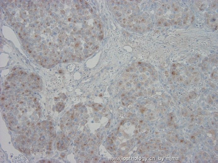
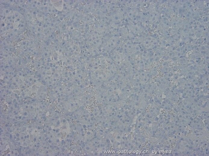
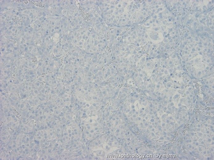
A few days later, a vertebral bone destructive lesion compressing the spinal cord was removed during decompression surgery, and photos 1~3 below were taken from the lesion. Multiple immunohistochemical stains were done, some of which were presented here: Figure 4 - AE1, 5 - Cam5.2, 6 - EMA, 7 - HepPar-1, 8 - alpha-fetoprotein, 9 - S100 protein, 10 - Melan-A, 11 - chromogranin A, 12 - neuron-specific enolase (NSE), 13 - synaptophysin, 14 - GFAP, and 15 - inhibin. What do you think?
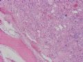 图1
图1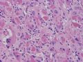 图2
图2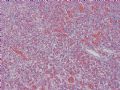 图3
图3 图4
图4 图5
图5 图6
图6 图7
图7 图8
图8 图9
图9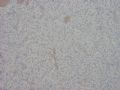 图10
图10 图11
图11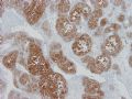 图12
图12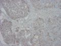 图13
图13 图14
图14 图15
图15
聞道有先後,術業有專攻
-
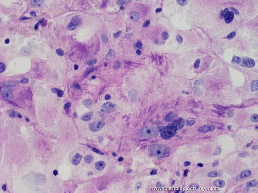
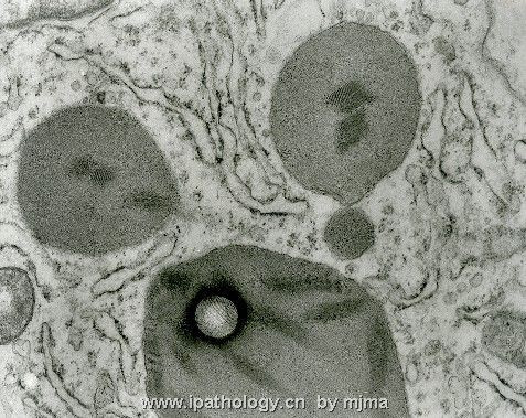
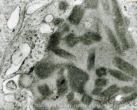
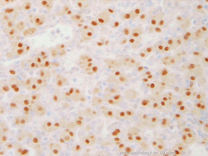
Figures 1~2 are from PAS stain after diastase pretreatment, figures 3~4 are electron micrographs taken from neoplastic cells, and figure 5 is an immunohistochemical stain of neoplastic cells using an antibody against TFE3 (transcription factor 3). These additional information led to the definitive diagnosis, which will be announced shortly. What do you think this is?
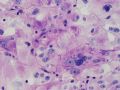 图1
图1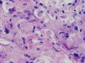 图2
图2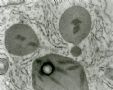 图3
图3 图4
图4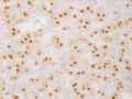 图5
图5
聞道有先後,術業有專攻
-
liguoxia71 离线

- 帖子:4174
- 粉蓝豆:3122
- 经验:4677
- 注册时间:2007-04-01
- 加关注 | 发消息
This is indeed a rare case of alveolar soft part sarcoma with wide spread metastases in liver, lung, bone and brain. It was extremely educational to me, and I wish to share what I learned with you.
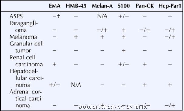
The initial brain tumor removed from this young man generated a long list of differential diagnoses of metastatic nature (no primary brain tumor has such histopathology). Those I considered seriously are (1) renal cell carcinoma (chromophobe variant of high nuclear grade), (2) hepatocellular carcinoma and hepatoblastoma, (3) Leydig cell tumor of testicular origin, (4) paraganglioma or pheochromocytoma, (5) oncocytic carcinoma of thyroid gland, salivary gland, or pancreatic origin, and (6) melanoma. Extensive immunohistochemical staining showed mild to moderate immunoreactivity to synaptophysin and strong immunoreactivity to neuron-specific enolase (NSE). Except for paraganglioma/pheochromocytoma, all of my other differential diagnoses were ruled out. After I exhausted my wisdon, I sent the case to a friend of mine, who told me the correct diagnosis after looking at the slides for 10 minutes. I stained it for PAS with diastase pretreatment, and saw NO CYTOPLASMIC PAS-POSITIVE CRYSTALS (the photos of PAS-positive crystals shown above were taken from the vertebral tumor resected later). Diagnostic electron microscopy and TFE3 immunostain were subsequently done and the diagnosis was confirmed.
Alveolar soft part sarcoma is rare. It often occurs in the extremities of young males. It is a highly aggressive malignancy of unclear histogenesis. Patients often present with symptoms related to metastastatic disease (seizure or weakness from CNS mets and pain from bone mets are most common) while the primary tumor remains either undetected or appears indolent (as is this case) at the time of presentation. Advanced stage (widespread metastases above and below diaphragm). After the suspicion of metastatic alveolar soft part sarcoma was raised based on the removed brain tumor, the patient was questioned and he reported a "leg mas" that had been present for a few months. Physical examination revealed a deeply seated, large soft tissue mass in his left upper calf. MRI showed this heterogeneously enhancing mass to be about 8 x 6 x 6 cm in size, and was in the muscles and fascia. Fine needle aspiration revealed neoplastic cells similar to those found in the brain (and subsequently, in the vertebral tumor).
It would not be necessary for me to copy what textbooks write about this fascinating malignancy here. I would encourage you read about the disease in updated textbooks or recent journals. A good summary by Weiss appeared 5 years ago in the American Journal of Pathology. It is available online at http://ajp.amjpathol.org/cgi/content/full/160/4/1197. A more recent review article by Folpe and Deyrup in 2006 is also available online at http://jcp.bmj.com/cgi/content/full/59/11/1127.
To summarize my lessons learned from this case: (1) never undervalue a complete history taking and physical examination, (2) PAS can be negative for alveolar soft part sarcoma, and (3) recognize my limitations and respect/welcome a second opinion from a colleague. I hope I will never miss this diagnosis again.
聞道有先後,術業有專攻
-
本帖最后由 于 2007-06-23 16:43:00 编辑
马老师的这例是一个深刻的有趣的病例,听听他讲的过程:
This is indeed a rare case of alveolar soft part sarcoma with wide spread metastases in liver, lung, bone and brain.本例的确是一罕见的软组织腺泡状肉瘤扩散转移到肝、肺、骨和脑部。 It was extremely educational to me, and I wish to share what I learned with you.给了我一个深刻的教训,我希望与大家分亨。
The initial brain tumor removed from this young man generated a long list of differential diagnoses of metastatic nature (no primary brain tumor has such histopathology).患者是一年轻男士,先以脑肿瘤切除术,我列出一长串的病理组织学的鉴别诊断(没有一个脑的原发瘤是如此的形态),Those I considered seriously are (1) renal cell carcinoma (chromophobe variant of high nuclear grade), (2) hepatocellular carcinoma and hepatoblastoma, (3) Leydig cell tumor of testicular origin, (4) paraganglioma or pheochromocytoma, (5) oncocytic carcinoma of thyroid gland, salivary gland, or pancreatic origin, and (6) melanoma. 我考虑了许多肿瘤如:1、肾细胞癌(高级别的嫌色细胞癌变异型),2、肝细胞癌和肝母细胞瘤,3、睾丸的Leydig 细胞瘤,4、副节瘤或嗜铬细胞瘤,5、甲状腺、涎腺或胰腺的嗜酸细胞癌,6、恶性黑色素瘤。Extensive immunohistochemical staining showed mild to moderate immunoreactivity to synaptophysin and strong immunoreactivity to neuron-specific enolase (NSE). 免疫组化显示:synaptophysin 弱到中度阳性,NSE强阳性,Except for paraganglioma/pheochromocytoma, all of my other differential diagnoses were ruled out.除了副节瘤和嗜铬细胞瘤之外,其它的需要鉴别的肿瘤都被一一排除。 After I exhausted my wisdon, I sent the case to a friend of mine, who told me the correct diagnosis after looking at the slides for 10 minutes. 穷尽我的智慧之后,送切片给我的一位朋友,他只是看了10分钟就确诊了。I stained it for PAS with diastase pretreatment, and saw NO CYTOPLASMIC PAS-POSITIVE CRYSTALS (the photos of PAS-positive crystals shown above were taken from the vertebral tumor resected later). 我经淀粉酶消化后 染PAS,没有看到胞浆内的PAS阳性结晶(PAS阳性的图象是椎骨肿物后染的阳性)。Diagnostic electron microscopy and TFE3 immunostain were subsequently done and the diagnosis was confirmed. 后经电镜和TFE3免疫组化确立诊断。
Alveolar soft part sarcoma is rare. 软组织腺泡状肉瘤是非常罕见的。It often occurs in the extremities of young males.常发生在青年男性的四肢, It is a highly aggressive malignancy of unclear histogenesis. 组织学起源末定,但有非常高的侵袭性。Patients often present with symptoms related to metastastatic disease (seizure or weakness from CNS mets and pain from bone mets are most common) while the primary tumor remains either undetected or appears indolent (as is this case) at the time of presentation.患者常因转移出现症状,如转移至中枢神经系统可出现头晕,无力。转移至骨胳可出现骨胳的疼痛,出现转移症状时往往还没有发现原发瘤(如本例)。 Advanced stage (widespread metastases above and below diaphragm).晚期可广泛转移至横膈膜的上下, After the suspicion of metastatic alveolar soft part sarcoma was raised based on the removed brain tumor, the patient was questioned and he reported a "leg mas" that had been present for a few months.脑部肿瘤被怀疑软组织腺泡状肉瘤后,询问患者几个月前,曾经有过腿部肿物史。 Physical examination revealed a deeply seated, large soft tissue mass in his left upper calf.检查患者发现左小腿腓肠肌上段深部有一大的肿块。 MRI showed this heterogeneously enhancing mass to be about 8 x 6 x
It would not be necessary for me to copy what textbooks write about this fascinating malignancy here.我有必要摘录一段教课书重新复习一下这个容易迷惑的肿瘤。 I would encourage you read about the disease in updated textbooks or recent journals. 我鼓励大家去读新版的教课书和以下的杂志,A good summary by Weiss appeared 5 years ago in the American Journal of Pathology. It is available online at http://ajp.amjpathol.org/cgi/content/full/160/4/1197. A more recent review article by Folpe and Deyrup in 2006 is also available online at http://jcp.bmj.com/cgi/content/full/59/11/1127.
To summarize my lessons learned from this case: (1) never undervalue a complete history taking and physical examination, (2) PAS can be negative for alveolar soft part sarcoma, and (3) recognize my limitations and respect/welcome a second opinion from a colleague. I hope I will never miss this diagnosis again.小结如下:1、不能忽视采积完整病史和查体,2、软组织腺泡状肉瘤的PAS可以是阴性的。3、认识到个人的能力是有局限性的,有必要欢迎同行提出不同意见。我希望今后再不要误诊这类病变。
-
wangliping 离线
- 帖子:139
- 粉蓝豆:1
- 经验:438
- 注册时间:2008-01-01
- 加关注 | 发消息


