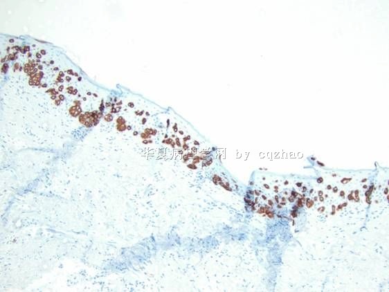
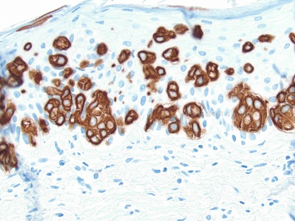
| 图片: | |
|---|---|
| 名称: | |
| 描述: | |
- 外阴Paget病和EMPD简要总结介绍(cqz-2)
-
本帖最后由 于 2009-08-23 04:51:00 编辑
Above summary is from one of our first year pathology resident. In fact she is resident only for the two month (starting from July, 2009).Residents and fellows need to prepare a lot of talks, case discussion, paper review in department conference, tumor board. They learn pathology knowledge when they prepare the talk. In fact I learned some new information from above our resident's presentaion. Also pathologists need to give reisdents and fellow many teaching course.
Some of the number or percentage may be not very accurate depending the papers you read. You can get a general information about EMPD.
I wish our young pathologists to learn from this case not nly the dx of Paget's. The more importance is to learn the way or thought, priniciple how to make your dx in clinical practice.
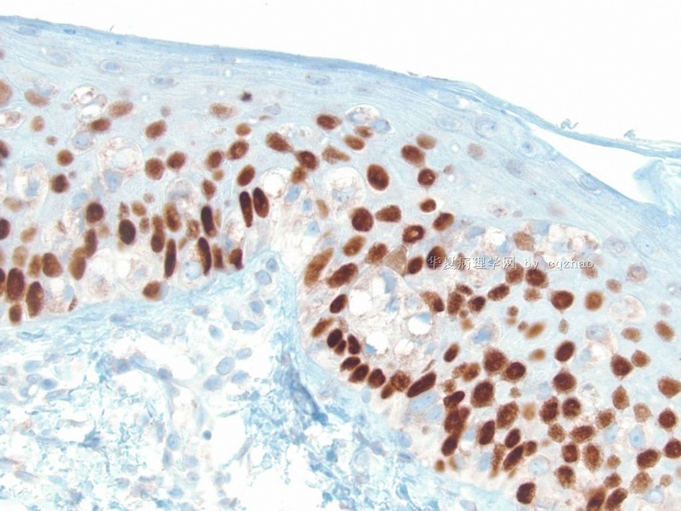
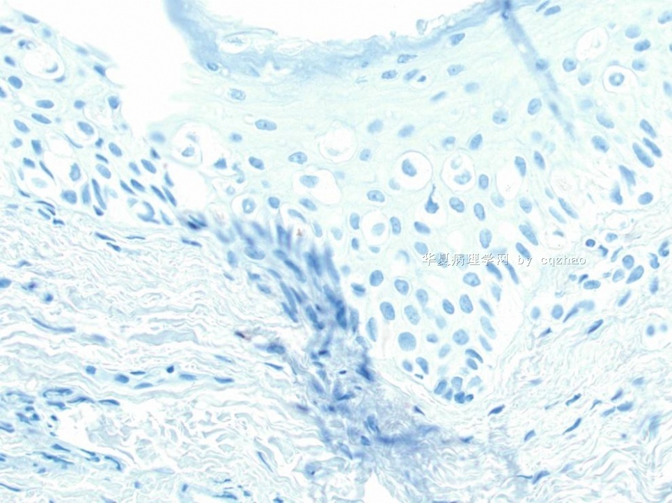
As pathologists we always should have differential dx for our cases even for some easy cases. For all paget's dieases we always order some stains to support our dx even though we almost are sure the dx based on the H@E slides. We do not want to misdiagosed melanoma or paget's. This is called low level but too significant mistakes. We may lost the job if we make this kind of mistakes.
Ok, I will finish this topic. Thank all of you who read or wrote the discussion for this topic. I especially appreciate 青青子矜 and Dr. Zhang for sharing their study or review summary.
It is the morning in China and in the US we are in the Friday night. I am playing 麻将game with my family tonight. I just learn it recently. I feel it is interesting and a good relaxing game.
Good weekend, cz
Conclusion
n Extramammary Paget’s Disease is an uncommon neoplasm, occurring most frequently in postmenopausal women on the vulva
n It was once believed to be secondary to underlying neoplasms, like mammary PD.
n Currently it is thought to be divided into two separate types, primary and secondary.
n IHC can aid in determining the type of EMPD and the origin of secondary EMPD.
n Toker cells may be the precursor cells in primary EMPD.
Is Toker cells Precursor Cells???
n Toker cells are a feature of the interface between the epidermis and the epithelium of lactiferous ducts of the breast, ectopic breast tissue and MLG.
n Morphologically appear to be benign counterpart of malignant cells in PD.
n Supported by IHC staining
-
本帖最后由 于 2009-08-22 08:03:00 编辑
Mammary-like glands (MLG)
n Glands that normally occur in the interlabial sulcus between the labia minora and labia majora
n Histologic features of female breast and sweat glands
n A, Opening of MLG with formation of duct-like structures by hyperplastic Toker cells (H&E).
B, CK7 positivity in hyperplastic Toker cells (CK7).From: Willman: Am J Dermatopathol, Volume 27(3).June 2005.185-188
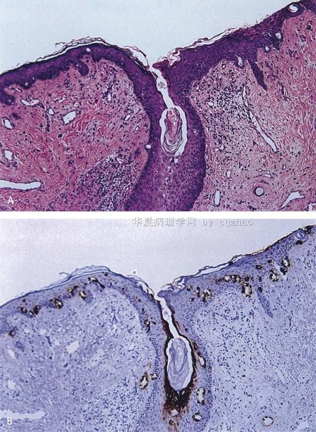
名称:图1
描述:图1
Primary EMPD
n Originate in the skin or apocrine sweat glands
n Not associated with distant adenocarcinoma
n Limited to the epidermis but may progress to invasive tumor, spreading to dermis, blood and lymphatics and potentially lethal metastases.
? Undifferentiated pluripotent cell of the epidermis or adnexa
? Toker cells of the vulvar epidermis
What are Toker cells?
n Intraepithelial cells with pale staining cytoplasm and bland cytologic features
n Located in the epidermis immediately adjacent to lactiferous ducts
n Considered a primitive germ cell of the lactiferous duct or inter-epithelial extension of lactiferous duct cells
n 10% normal nipples on H&E
83% normal nipples and 65% of accessory nipples with CK7 IHC
-
本帖最后由 于 2009-08-22 18:54:00 编辑
Glad to see above from Dr. Zhang and 青青子矜. I am a gynecologic/breast pathologist, but never do any research or write review about Paget's. Both of you did some research or wrote review articles in this area, so you know more detailed information. Thank for your expert's in-put.
I will continue to complete our resident's summary.
-
本帖最后由 于 2009-08-22 07:51:00 编辑
Pathogenesis of EMPD
n Unlike MPD, EMPD does not have a strong association with underlying adenocarcinoma (92-100% vs ~25%)
n Frequency of association of EMPD with underlying neoplasm varies with the site of the disease
n Two schools of thought:
n Paget's disease must arise from underlying neoplasm
n Paget's disease arises primarily in the epidermis
n Current theory is that there is dual origin of EMPDàPrimary vs. secondary
n Most cases of EMPD are primary
Two distinct types of EMPD
Type I- endodermal phenotype; associated with distant carcinomas, secondary
Common IHC phenotype
Type II- ectodermal/cutaneous; primary
-
本帖最后由 于 2009-08-22 07:48:00 编辑
Pathologic Features of PD
n Invasion of the epidermis by Paget cells (PC)
n Epithelial cells with abundant, clear cytoplasm
n Large, centrally situated nuclei with atypia and pleomorphism
n Signet ring aspect due to intracytoplasmic sialomucin
+ PAS, mucicarmine,
90% vs only 40% MPG
Pathologic Features of Extramammary PD
Lower epidermal layers, occasionally forming gland like structures with a central lumen
Outer epithelial layers of hair follicles or sweat gland excretory ductsEdematous dermis with dilated blood vessels and polymorphous inflammatory cell infiltrate
-
本帖最后由 于 2009-08-22 07:43:00 编辑
Vulvar EMPD
n 65% cases of EMPD
n Most cases are primary
n 4-17% associated with underlying adnexal neoplasm
n 11-20% associated with distant carcinoma
n Endometrial, endocervical, vaginal, bladder, colon, rectum, ovary, liver, gallbladder, skin
Differential Diagnosis
n Melanoma
n Bowen's disease
n Seborrheic or contact dermatitis
n Superficial fungal infections
n Basal cell carcinoma
n Flexural psoriasis
n Lichen sclerosus
-
本帖最后由 于 2009-08-22 07:46:00 编辑
Extramammary Paget's Disease (EMPD)
Epidemiology
n More rare than mammary Paget抯 disease (MPD)
n accounting for 6.5% of all cases
n MPD account for 1-4% of breast cancer
n 65-70 years of age, 90% over 50
n Female predominance of the vulvar localization (2% or less of primary vulvar neoplasms)
n No definite proof of a genetic predisposition
Clinical Features
n Insidious onset
n Pruritus
n Well-demarcated plaques may be separated by clinically normal skin that is pathologically involved
n Rarely, hypopigmented/hyperpigmented macules
n "underpants pattern erythema"due to lymphatic invasion and regional node enlargement and rapidly fatal distant metastases
n Diagnosis is often delayed up to 2 years until histological examination is performed secondary to chronic dermatosis not responding to local treatments.
Topographic Forms
n Perianal-20% EMPD
n Affects men and women equally, 63 yo
n Associated with adenocarcinoma of anus and rectum (~75%)
n Male Genitalia-14%
n Associated with underlying carcinoma of the ureter, bladder, prostate, testicle (11%)
n Axillary-more frequent in men, unilateral
n Exclusion of breast neoplasm is mandatory
n Ectopic-skin areas devoid of apocrine glands
n Chest, arms, fingers, eyelids, cheeks, scalp
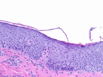
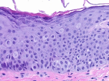
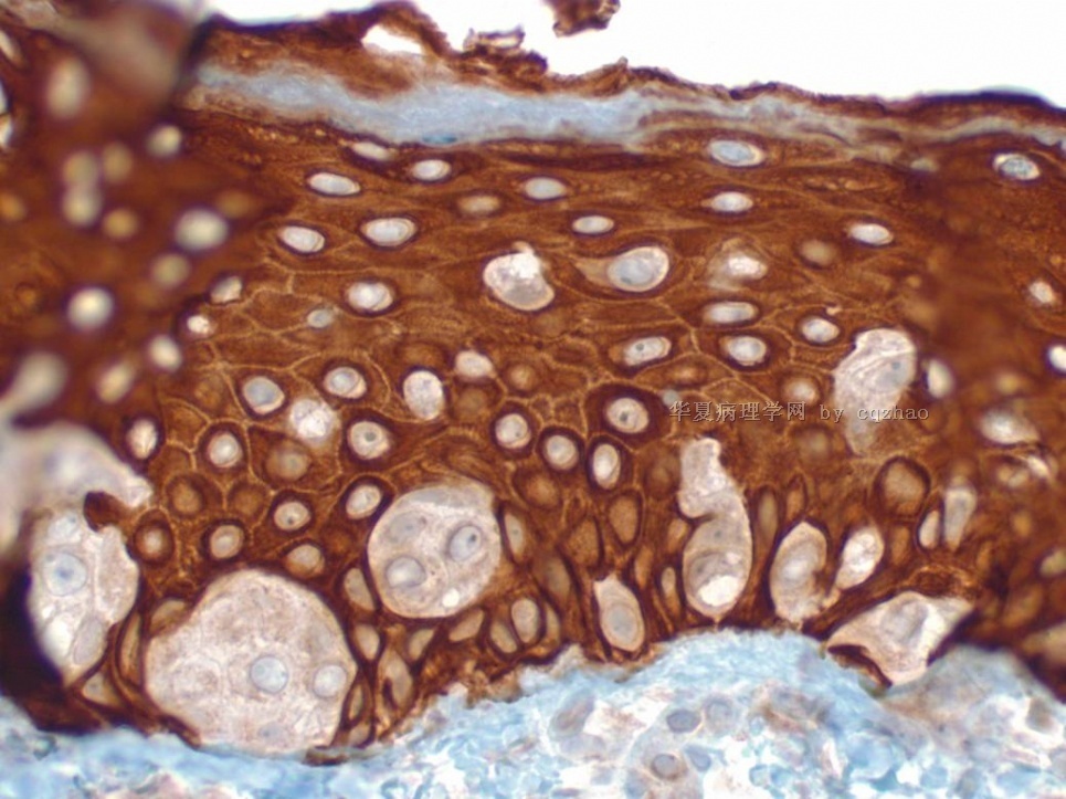
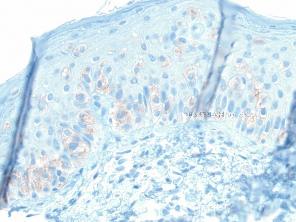
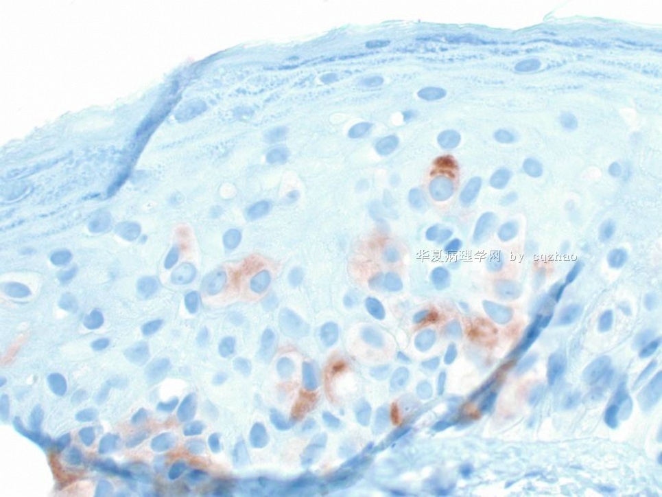
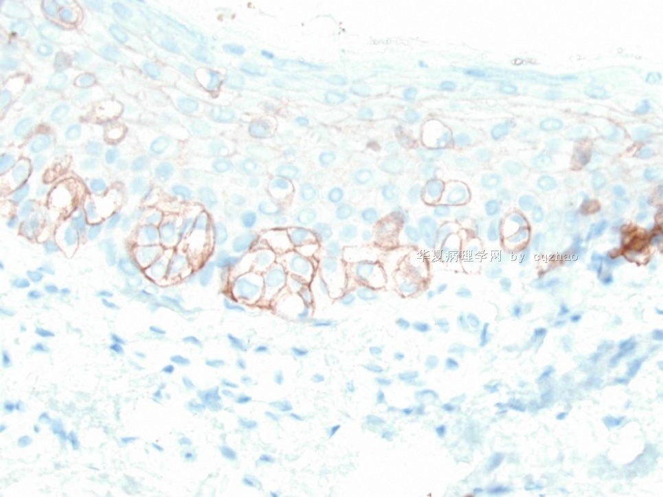
This is a vulvar Paget's/primary Paget's or called Extramammary Paget’s Disease
One of our first year residents made some summary about Paget's and showed in our morning study conference. I pasted the summary in the following.
As pathologists we should know lesions in more detailed. It is not enough to make dx only.
