| 图片: | |
|---|---|
| 名称: | |
| 描述: | |
- 外阴赘生物
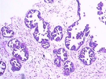
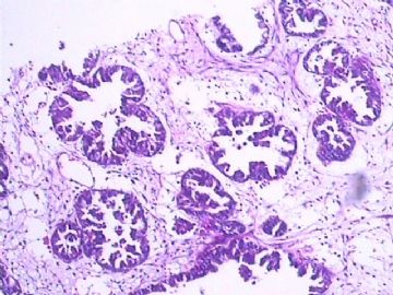
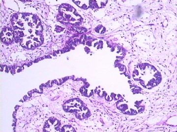
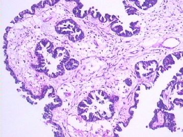
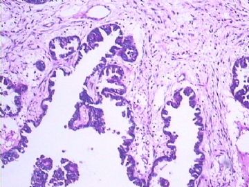
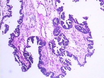
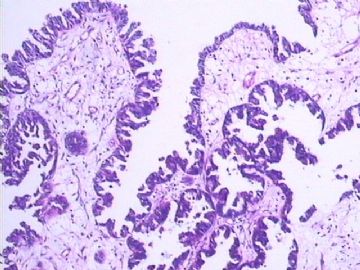
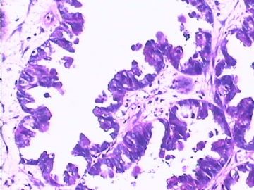
Please provide couple low power photos of the lesion. My immediate impression is a benign lesion. Top differentials will include an ectopic mammary glands and bartholin gland cyst. A large dilated glands with surrounding small glands indicates a lobular architecture which can be seen in both lesion. Bartholin gland cyst consist often of blend mucinous glands. This one is less mucinous to me. For ectopic breast tissue, you need to search if there is a myoepithelial cell layers beneath the glandular epithelial cells. Please search for that and give us couple high power view of the glandular cells/layers.
I will be very very careful to jump on "adenocarcinoma" for this tiny vulvar lesion with so blend morphology in this 28 year old young woman before ruling out benign lesions!

- 不坠青云之志,长怀赤子之心
-
dingcaixia 离线
- 帖子:40
- 粉蓝豆:1
- 经验:180
- 注册时间:2009-08-04
- 加关注 | 发消息
-
dingcaixia 离线
- 帖子:40
- 粉蓝豆:1
- 经验:180
- 注册时间:2009-08-04
- 加关注 | 发消息
杨斌老师意见:
请提供点此例的低倍图片。我的第一印象是个良性病例。首先想到的鉴别诊断是异位乳腺和巴氏腺囊肿。 一个大的腺体周边环绕一些小腺体提示是一个小叶状结构,而这种结构在上面两种病例中都可能看到。巴氏腺囊肿一般由混合型粘液性腺体构成。此例好像没看到粘液。 如果是异位乳腺组织,就需要在腺上皮细胞下找一下有没有肌上皮细胞层。请仔细找一下,提供点这样的区域的高倍图像。28岁女性,外阴如此之小的肿物,形态学表现为混合性,在排除良性病变之前我会极为小心,尽量先不发腺癌

- 赚点散碎银子养家,乐呵呵的穿衣吃饭

