| 图片: | |
|---|---|
| 名称: | |
| 描述: | |
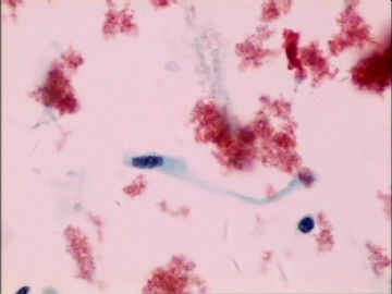
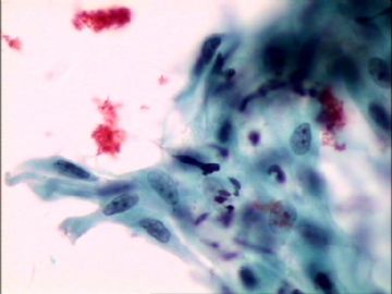
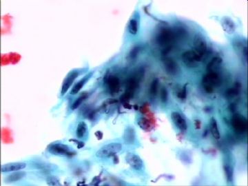
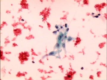
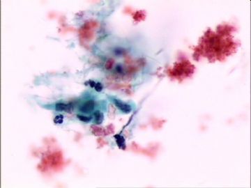
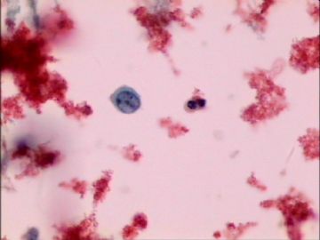
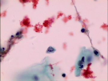
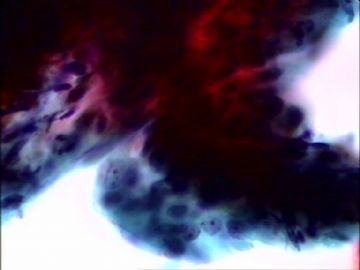
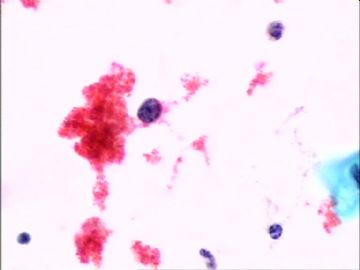
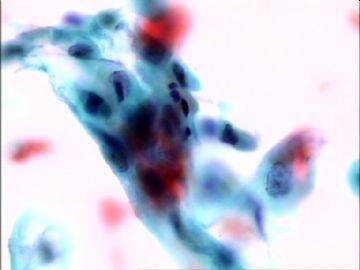
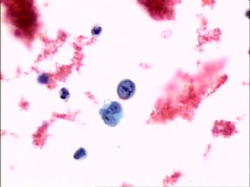
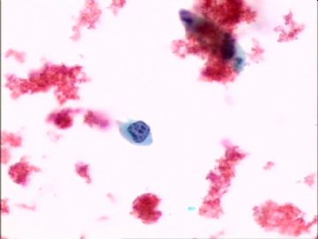
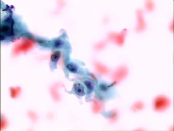
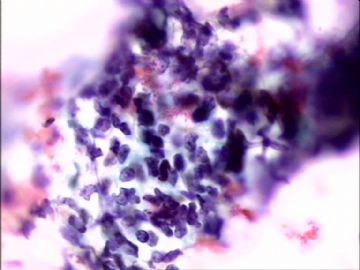
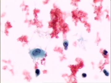
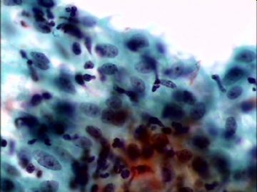
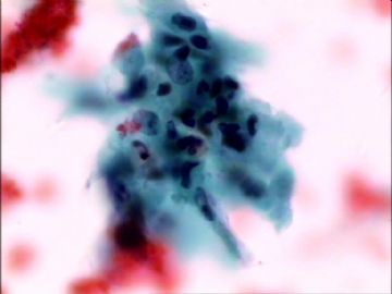
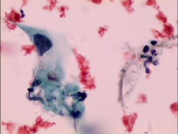
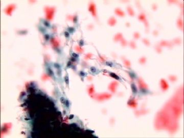
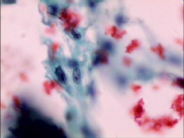
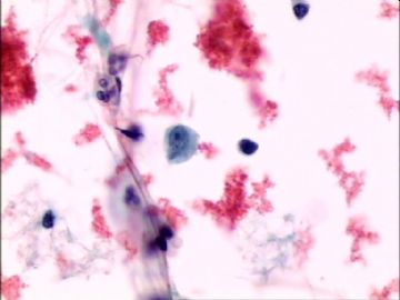
- 液基.65岁,绝经十余年,阴道不规则流血2天
-
本帖最后由 于 2009-08-02 21:34:00 编辑
Firstly I need to mention that what we discuss here is subjective because our impression is based on some photos only. 首先我需要提及的是我们在这里讨论的主观性,因为我们仅仅基于一些图片。
Agree with above analysis. It is abnormal Pap. I suggest to call ASC-H, and AGC. The reasons are what you have described. The key is that the women should get tissue sampling.同意上面的分析。这是一个不正常的巴氏涂片。我建议报ASC-H和AGC;这些原因正如你描述的。关键是这个患者应该进一步组织活检。
Remember three things: 请记住三点
1) women should have endocervical and endometrial sampling even we call AGC, endocervical for women >or =35 years..1、在35岁及以上的女性,我们报AGC后应该进行宫颈管和宫内膜活检。
2) 70- 80% women with AGC will not have lesions in the follow-up histologic examination. As pathologists or cytopathologists we feel bad for ourselves. But this is the limitation of pap test. What can we do? 2、70-80%的AGC女性在组织学随访中是阴性的。作为病理学家或细胞病理学家感到难过;但是这是巴氏的局限性。那我们能做什么呢?
3) Pap test is a screening test (principle). If you want to call malignant in Pap test you must be100% sure of this diagnosis. Otherwise, please use atypical, or at most favor or suspicious ....words. Bottomline is that you need to havecomminication with the clinician to express your worry for this case and suggest women to have bx.
3、巴氏试验是筛查试验(原则)。如果你想在巴氏中报恶性肿瘤,你就得有100%的把握肯定这个诊断。否则,请用非典型性、或可能性大或可疑等等字眼。低线是你必须跟临床医生沟通表明你对这个病例的担心和建议必须进行活检。
If you spend time to consider it is malignant or atypical, I suggest you call atypical.如果你花精力考虑这是恶性或非典型性,我建议你叫非典型性。
cz
掌心0164翻译。
-
本帖最后由 于 2009-08-03 20:56:00 编辑
1. AGC or AGC, endocervical cells need to have endometrial sampling (if age 35 or older/>35 y or at risk for endometrial neoplasia) and colposcopy. High risk HPV testing is also suggested to these women.1、 AGC或AGC宫颈管来源,需要进行阴道镜下颈管活检和宫内膜活检(如果年龄在35岁及以上,宫内膜瘤变风险增加),这些女性也可建议进行高危HPV检测。
2. Atypical endometrial cells: endometrilal and endocervical sampling. If no endometrial lesion noted, colposcopy is suggested.非典型宫内膜细胞:内膜和颈管活检;如果没有发现内膜病变;建议阴道镜。
Above are ASCCP (American Society of Colposcopy and Cervical Pathology) guideline.
我看到的资料好像说是>=40岁. I do not know what book or paper you read. Or Chinese society has a guideline.上述是ASCCP(美国阴道镜和宫颈病理学会)指导准则。
Please read ASCCP guideline if you are interested如果你感兴趣请读ASCCP(美国阴道镜和宫颈病理学会)指导准则:如下
http://www.ipathology.cn/forum/forum_display.asp?keyno=124776
-
本帖最后由 于 2009-08-01 19:58:00 编辑
巴兄您真看得起我呀;这个病例我还没有找到一个合适的诊断来解释图片中的现象。
1、单个高核浆比、胞浆稀少、染色透亮和核仁明显的细胞(1图右下、6、7、9、11、12、13、15图)是同一种细胞吗?如果说12、13、15是上皮来源比较肯定之外;其他的尤其9和11的细胞是上皮来源吗?怎么象间质来源呢?
2、成团的细胞来源上皮(除14图的裸核外)问题不大;象修复性的病变;结构紊乱;核大小不等;大核仁。尤其第8图深染色不透光;难以看清楚;但是下面提示象腺来源;不敢肯定。
3、鲜血的背景,不象肿瘤坏死的背景;取材所导致还是病变位置比较远导致的出血残留呢?
4、临床病史也支持恶性诊断
如果是我自己现实的病例我就请上级医生会诊。这里我就冒失猜一个诊断、一点底都没有。
诊断考虑如下:
查见恶性肿瘤细胞:癌可能性大不排除合并肉瘤。请临床提供盘腔B超、既忘病史等等。请临床进一步处理。
最后肯定各位老师给予指正,先谢谢了。

- 掌心0164
啊,这个病例的确很棘手,很难给个恰当的诊断。图4、6、12、13这些有个小核仁核染色轻淡的我倾向是化生的鳞,至多是不典型修复,图2、3团块的排列倒是很象腺,有飞出去的感觉,可报AGC。。。需要更多的临床资料。“65岁老年女性有阴道出血”,这样不明确的图象,如果实际工作中我碰到的话,我会找临床医生沟通一下或请病人来科里看病历并询问详细病史,出血程度?出血多的话临床不管TCT结果就直接是分段诊刮的指征啦。妇科检查的宫颈情况?既往病史?等等。。。最后可能给个描述性诊断,建议由临床医生结合具体情况再做下一步治疗处理。
期待后续报道
