| 图片: | |
|---|---|
| 名称: | |
| 描述: | |
- Rectus mass FNA
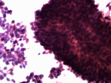
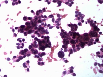
This is a 62-year-old man has history of 3 carcinomas: 2006 high-grade urothelial carcinoma (transitional cell carcinoma), 2008 papillary thyroid carcinoma, 2009 prostate carcinoma (on prostatectomy it showed gleason 5, 4, and 3 patterns). He had cystectomy, prostatectomy, and thyroidectomy. This FNA was done under the CT, it is positive for malignancy. Can you pick which primary carcinoma now is metastasized?
This should be a easy case. Please just look at the morphology carefully.
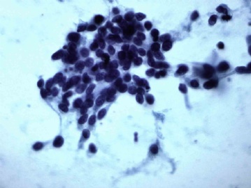
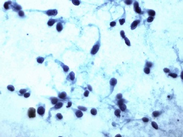
I know that a lot of you like the H&E stain, the first 2 photos are H&E stain, the last 2 are Pap stain, I hope you can appreciate the difference.
LjmLjm, Liguoxia71, and 江边观潮人, you are all right on the diagnosis. LjmLjm mentioned 蝌蚪样细胞 (cercariform cells) is the reason that I posted this case. I am pasting the original paper abstract by Dr. Powers describing this kind of cells:
"Cercariform" cells: a clue to the cytodiagnosis of transitional cell origin of metastatic neoplasms?
Diagnostic Cytopathology, 1995, 13:15-21.
Powers CN, Elbadawi A.
Department of Pathology, State University of New York, Health Science Center, Syracuse 13210, USA.
The "cercariform" cell is described as a distinct cytomorphologic clue that may be helpful in the diagnosis of metastatic transitional cell neoplasms, particularly low grade. This cell has a nucleated globular body and a cytoplasmic process with a nontapering, flattened, bulbous or fishtail-like end. The cercariform cell corresponds to intermediate cells in histologic and ultrastructural preparations of normal urothelium. The cercariform appearance is the result of pseudostratification of both normal and low-grade neoplastic urothelium. The unique features of cercariform cells make them readily distinguishable from neoplastic squamous cells as well as spindle cells of mesenchymal origin.


