| 图片: | |
|---|---|
| 名称: | |
| 描述: | |
- 肝脏巨大肿瘤
-
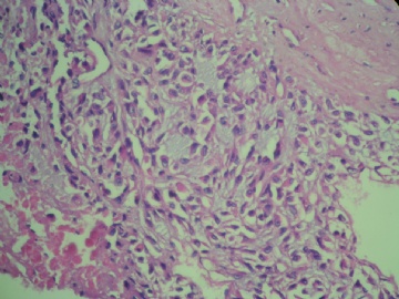
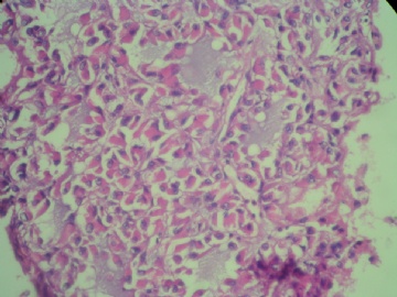
Amazing case this is. I would agree that the lesion is a malignant neoplasm, but its classification behooves me. Neoplastic cells are hepatoid with large vesicular nuclei, prominent nucleoli, and plump polygonal eosinophilic cytoplasm. Ther are placed in a environment rich in extracellular myxoid substance, and do not form the usual rosette/pseudoglandular, pelioid, trabecular or plate-like architecture with sinusoid formation expected in classic hepatocellular carcinoma. The relatively slow progression (5 years without treatment) also argues against classic hepaticellular carcinoma. Fibrolamellar variant of hepatocellular carcinoma may be this slow growing and indolent, but patients are usually younger. I think one needs immunohistochemistry to help classify this interesting malignancy. I would start with HepPar-1, alpha-fetoprotein, AE1, CEA, CK5/6, and calretinin to begin with. Looking forward to learning more from your on-going investigation.

聞道有先後,術業有專攻


