| 图片: | |
|---|---|
| 名称: | |
| 描述: | |
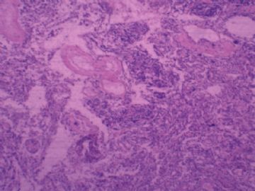
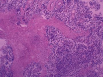
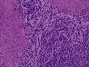
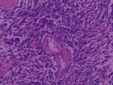
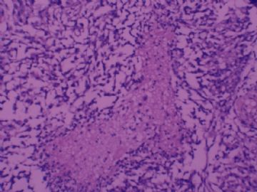
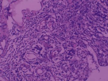
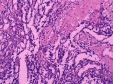
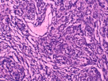
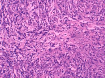
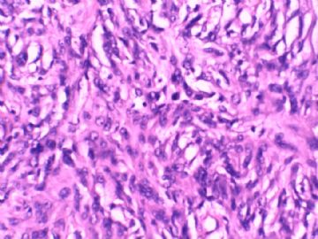
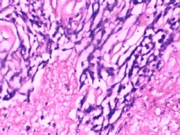
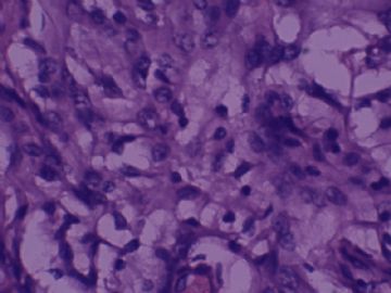
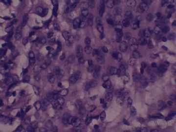
- 右额叶肿瘤,脑膜瘤?
-
本帖最后由 于 2009-07-16 16:15:00 编辑
Yes, I believe this is a case of meningioma. Suboptimal histology suggest chordoid meningioma, but I think most of the spaces between cells are artifacts rather than true intercellular mucin-rich spaces seen in chordoid meningioma. There appears to be multifocal tumor necrosis and, at least in a few better preserved areas, prominent nucleoli. These suggest that this may not be a grade I benign meningioma. One has to search and count mitotic figures carefully in better preserved areas, and look for the other atypical features - hypercellularity, small cells with high nucleocytoplasmic ratios, patternless growth, and brain parenchymal invasion. IMC for EMA, PR and MIB-1 may be very helpful.
liguoxia译:我认为该例是脑膜瘤。组织学图片欠佳,似在提示脊索样脑膜瘤,但是细胞之间的区域多数是人工假象,而不是真正的脊索样脑膜瘤中见到的细胞间富于粘液区域。图片显示了多灶性肿瘤坏死,核仁突出(至少在几个保存好一些的区域中能见到)。这些提示不会是良性脑膜瘤。应该在保存好的区域仔细寻找计数核分裂,寻找其它非典型特征:细胞密集、核浆比高的小细胞、无序生长方式、脑实质浸润。免疫组化EMA、PR、MIB-1会有帮助。

聞道有先後,術業有專攻
-
lfl001200546 离线
- 帖子:2808
- 粉蓝豆:40
- 经验:2808
- 注册时间:2007-02-14
- 加关注 | 发消息

