| 图片: | |
|---|---|
| 名称: | |
| 描述: | |
- NP (11)
| 姓 名: | ××× | 性别: | M | 年龄: | 77 |
| 标本名称: | Intracerebral hematoma and adjacent brain | ||||
| 简要病史: | Sudden onset of mental status change and found to have a right frontal lobe hematoma with mass effect and midline shift. He underwent emergent craniotomy for hematoma evacuation. | ||||
| 肉眼检查: | Fresh blood clot and associated gray-tan brain tissue | ||||
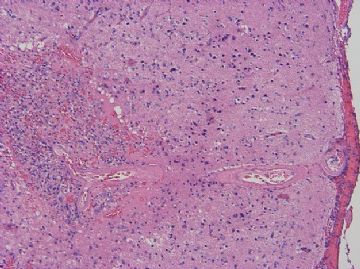
Figure 1 is cerebral cortex overlying hematoma (100x)
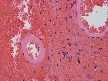
Figure 2 shows small leptomeningeal blood vessels in blood clot (400x)
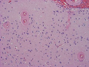
Figure 3 shows superficial cortex (200x)
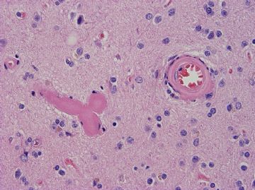
Figure 4 shows small cortical blood vessels (400x)
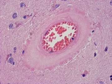
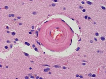
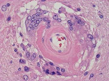
Figures 5-7 show cortical blood vessels (600x)
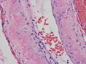
Figure 8 shows a large leptomeningeal blood vessel (400x)
What is your diagnosis? Is there any stain that may help you confirm the diagnosis?

聞道有先後,術業有專攻
-
本帖最后由 于 2009-07-16 15:57:00 编辑
This is indeed a classic case of "cerebral amyloid angiopathy." Classic histopathology is eosinophilic homogenization of thickened walls in leptomeningeal and intracortical small blood vessels. In severe or chronic cases, the amyloid deposits may appear as concentric rings (as seen in Figure 6). They may also rarely elicit phagocytic and foreign body giant cells reaction (as seen in Figure 7), which should not be confused with granulomatous meningoencephalitis. Larger vessels (Figure 8) may also be affected. The protein deposited is beta-amyloid peptides derived from beta-amyloid precursor protein, similar to the peptided found in senile plaques in neuropil of normal aging brains and Alzheimer disease brains. It is associated with increased risks for large lobar hemorrhage (as in this case) small cortical microinfarcts (Figure 1). Many (not all) Alzheimer disease brains have severe cerebral amyloid angiopathy, and many (not all) cases of cerebral amyloid angiopathy have concurrent Alzheimer disease. Conventional Congo red (red-green birefringence) and thioflavin-S (green fluorescence) stains can demonstrate amyloid in these deposits.
liguoxia71试译,请指正:本例是典型的大脑淀粉样血管病。其典型组织病理学表现为软脑膜和皮质内小血管壁增厚均质嗜红染。病变呈慢性或严重时淀粉沉积物同心圆状(如图6),甚至引起吞噬细胞、异物巨细胞反应(如图7),注意不要与肉芽肿性脑膜脑炎混淆。大血管也可以受累(图8)。沉积的蛋白是源自β-淀粉前体蛋白的β-淀粉多肽,类似于在正常老化大脑和阿尔茨海默病的大脑中神经毡、老年斑中发现的多肽。淀粉样血管病使大的叶性出血(如本例)、皮质小梗塞几率增加(图1)。许多阿尔茨海默病脑组织(不是所有病例)可见明显的大脑淀粉样血管病,许多大脑淀粉样血管病(不是所有病例)并发阿尔茨海默病。常规刚果红(红绿双折射)、硫磺素—S(绿荧光)染色可以显示沉积的淀粉样物。

聞道有先後,術業有專攻

