| 图片: | |
|---|---|
| 名称: | |
| 描述: | |
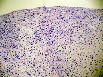
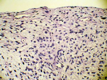
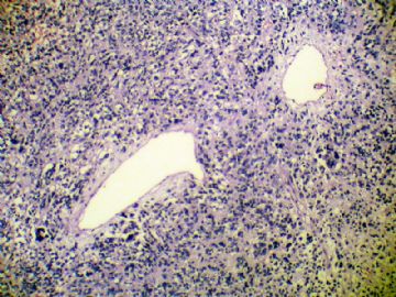
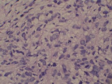
- 右侧脑室三角区占位
-
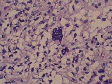
This tumor is located at the right lateral ventricular wall near the atrium, a location usually not supportive of dura-based meningioma. I wonder why did MRI show a tumor suspicious for meningioma at this location. The photos uploaded show no clearcut meningothelial differentiation. Instead, I see cytologic atypia and pleomorphism that suggest a high grade astrocytoma (at least WHO grade III). Are there mitotic figures, necrosis or vascular/endothelial proliferation? EMA, PR and GFAP immunohistochemical stains would help.

聞道有先後,術業有專攻

