| 图片: | |
|---|---|
| 名称: | |
| 描述: | |
- What stains are you using for FNA cytology?
From what I have learned and the interactions I had with our pathologist friend in China, I would like to bring this topic to discuss: What are the stains you routinely used for FNA cytology? H&E or Diff-Quick or Pap or any 2 of the above 3?
What are the advantages and disadvantages of each of the stains?
Thank you for the comments Urbino! I now posting some pictures of diff-quick for illustration:
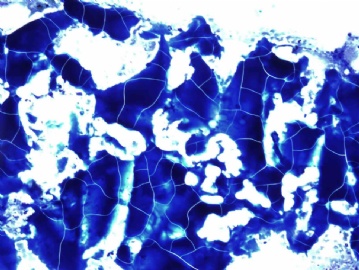
Picture 1: you can see the nice thick colloid with cracking artifacts, it is typical of colloid nodule.
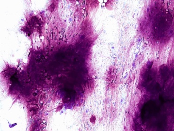
Picture 2: shows the typical chondroid-myxoid stroma of pleomorphic adenoma
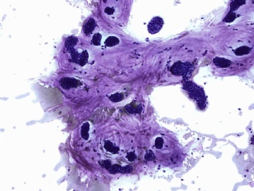
Picture 3: a case of breast colloid carcinoma, you can see the "balls" of tumor cells, the Diff-Quick stain also highlights the background mucin.
Is 亚甲蓝 toludine blue? Actually I had a chance to visit and learn FNA cytology at the University of California at San Francisco, their pathologists perform more than 3000 FNAs a year in their own FNA clinic. They used toludine blue for their rapid review and Pap stain for their permanent stain. I liked it a lot.
Let's further discuss the stain options we have. The reason that most of the cytopathologists in the US don't like the rapid H&E is that the stain introduces artifacts. For example, in thyroid FNA (very common specimen), it produces pseudo "hole" in the nucleus, which can lead the pathologist to over-diagnose papillary carcinoma. Also, it dissolves the colloid, which can lead the pathologist to over diagnose follicular neoplasm. One of the cases recently posted here by LjmLjm used H&E, although I don't see much colloid on the picture, the follicular cells are pretty much in flat sheet. some people immediately jump into "follicular neoplasm". It is true that Pap stain will dissolve colloid too, but Pap stain shows excellent nuclear cytomorphology and it is very close to H&E in term of cell size (for most of us doing both cytology and surgical pathology, it is helpful). Although not showing as clear a cytomorphology as Pap stain, the diff-quick (or it's variant) stain tends to preserve or highlight the background materials, like colloid, chondroid-myxoid stroma of pleomorphic adenoma, and extracellular mucin. With the combination of both Pap and diff-quick, it tends to give the cytopathologists enough weapons to attack the very challenging cases.
Thank you for all the discussions here, I would like to hear more opinions.
-
I need to thank Urbino for the very good comments. Let me give you an overview on this topic in the United States. I have visited many cytopathology labs in the country. Pap stain and Diff-Quick (or other kind of Romanowsky stain) are the most commonly used stains on FNA cytology. H&E is clearly much less used. One important fact is that if you submit a paper to any major journal of cytology in the US, if you just have the H&E stained pictures, your paper will have a hard time to get published. I would like to hear more discussions or comments regarding this topic. Thank you!


