| 图片: | |
|---|---|
| 名称: | |
| 描述: | |
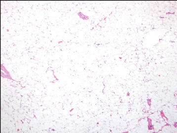
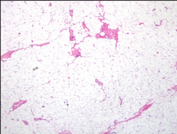
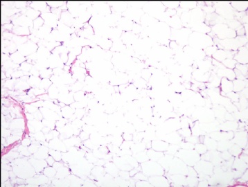
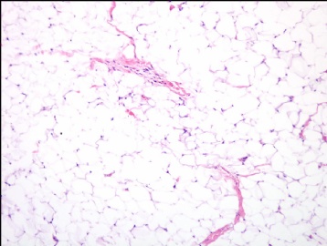
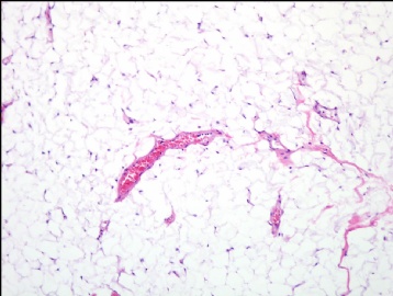
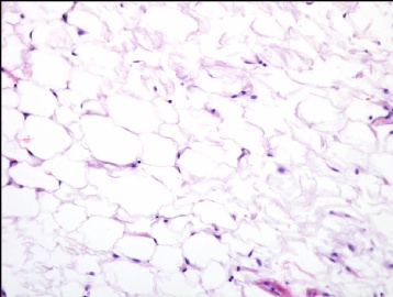
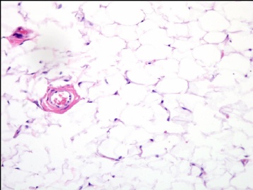
- B1638腹膜后肿物
-
链接里有关于高分化脂肉和脂肪瘤的10点要领,觉得蛮好,闲得无聊,翻译如下:
1. The discrimination between ordinary lipoma and atypical lipomatous tumor/well-differentiated liposarcoma (ALT/WDL) is a common diagnostic challenge in diagnostic soft tissue pathology.
普通型脂肪瘤和非典型/高分化脂肪肉瘤(ALT/WDL)的鉴别诊断是软组织病理学诊断工作中的一个难点。
2. Histologic, cytogenetic, and molecular genetic data support the idea that these two major groups of adipose tissue tumors are distinct biologic entities in spite of overlapping histologic features.
尽管有组织学特征上的交叉重叠部分,但是这两种肿瘤的组织学,细胞遗传学和分子遗传学数据都支持两者是不同的生物学疾病实体。
3. Careful tissue sampling and histologic examination are the major and simplest steps for the correct discrimination between a lipoma and an ALT/WDL.
为了正确鉴别出脂肪瘤和ALT/WDL,认真细致的组织取材和组织学观察是主要的,也是最简单有效的的措施。
4. Lipomas can be superficial or deep seated and are histologically characterized by mature fat cells with no cytologic atypia/hyperchromasia/pleomorphism. Occasionally, lipoblasts can be seen in lipomas, especially near blood vessels. However, they do not exhibit cytologic atypia/hyperchromasia/pleomorphism.
脂肪瘤表浅,或者位于深部,组织学上以成熟脂肪细胞为特征,细胞无异型/深染色质/多形性。少数情况下,脂肪母细胞也能在脂肪瘤中看到,特别是近血管区域,但是,它们不显示细胞异型/深染色质/多形性。
5. ALT/WDL are histologically characterized by the presence of mixed population of lipoblasts and lipocytes but more importantly by atypical hyperchromatic pleomorphic cells. These tend to be more commonly found near blood vessels and within fibrous septa. It is also important to note that the identification of lipoblasts is not a necessary criterion for the diagnosis of an ALT/WDL.
ALT/WDL组织学上以脂肪母细胞和脂肪细胞混合存在为特征,但更重要的特征是不典型性具有深染色质的多形性细胞。这些细胞一般很容易在血管周围和纤维间隔内找得到。另外要强调指出的是脂肪母细胞的辨认不是诊断ALT/WDL的必要标准。
6.The discrimination between lipoma and ALT/WDL becomes challenging when the diagnostic atypical hyperchromatic pleomorphic cells are rare or have subtle atypical cytologic features. Many cell types or tissue artifacts can simulate these cells, including lockhern artifact (intranuclear holes), activated fibroblasts/myofibroblasts, multinucleated giant cells, and degenerated skeletal muscle fibers. However, in most instances these cells can be correctly identified by careful examination. However, ALT/WDL cells with subtle cytologic features can be very difficult to recognize by histologic exam. In these situations, ancillary studies may be important to establish the correct diagnosis.
当诊断性异型深染色质多形细胞稀少或仅具有轻微细胞学异型时,脂肪瘤和ALT/WDL的鉴别变得很困难,许多细胞类型或一些组织假象中都可见类似细胞,如lockhern(核内空洞),活化的纤维母/肌纤维母细胞,多核巨细胞,和退变的骨骼肌纤维,大多数情况下,这些通过认真检阅都能正确分辨。但是只有轻微细胞异型特征的ALT/WDL仅仅通过组织学观察要鉴别出来相当的难,在这种情况下,要做出正确诊断要通过一些辅助手段。
7.Lipoma and ALT/WDL have very distinct cytogenetic and molecular genetic characteristics:
a.Lipomas are mainly characterized by rearrangements of chromosome 12q13~q15 with several partner chromosomes in approximately 50-60% of cases, especially chromosome 3 (figure 1). Lipomas without 12q13~q15 rearrangements frequently show rearrangements of chromosome 6p21. Several lipoma fusion genes have been identified and the most common is LPP-HMGA2, product of the t(3;12)(q27-q28;q14-a15). Interestingly, lipomas grow relatively well in culture and, in our experience, 60-70% of them show abnormal karyotypes.
b.ALT/WDL usually exhibit supernumerary ring or giant maker chromosomes by standard cytogenetic analysis (figure 2). These abnormal chromosomes are composed by amplified genomic material mainly derived from chromosome bands 12q13~q15. These bands contain several cancer genes, including MDM2, SAS, CDK4 and HMGA2. MDM2 seems to be the most consistently amplified gene in ALT/WDL (>99% of cases). Amplification of these genes is not observed in lipoma.
脂肪瘤和ALT/WDL具有完全不同的细胞和分子遗传学特征:
a.50%-60%的脂肪瘤,以染色体12q13~q15区与一些染色体,尤其是与3号染色体的重排为主要特征(图1)。无12q13~q15重排的脂肪瘤常常显示染色体6p21的重排。一些脂肪瘤融合基因也被证实,最常见的是LPP-HMGA2, t(3;12)(q27-q28;q14-a15)的产物,有趣的是,脂肪瘤在体外培养生产相对良好,60-70%显示异常核型。
b.ALT/WDL常常显示大量的环状和巨大染色体(图2),这些异常染色体是由源于染色体12q13~q15带的扩增基因序列组成的,这些扩增带常含有一些癌基因,包括MDM2, SAS, CDK4 and HMGA2。MDM2似乎是ALT/WDL中最恒定的扩增基因(超过99%病例)。脂肪瘤没有这些基因扩增。
8.In the clinical practice several ancillary techniques can be used to discriminate lipomas from ALT/WDL: standard cytogenetics, molecular cytogenetics (fluorescence in situ hybridization and chromogenic in situ hybridization), molecular genetics (polymerase chain reaction) and immunohistochemistry. Table 1, which is not intended to be comprehensive, summarizes some of the advantages and disadvantages for each of them (key papers are also listed in the suggested references).
当前的辅助检测:标准细胞遗传学,分子细胞遗传学项目(荧光原位杂交FISH,显色原位杂交CISH),分子遗传学(多聚酶链反应PCR),免疫组化。表1比较了优缺点。
Table 1. Advantages and disadvantages of ancillary techniques for the discrimination between lipoma and ALT/WDL (表略)
9.And why this discrimination is important? This is a very important question that has not been fully answered but the overall experience is that lipomas have a lower local recurrence that ALT/WDL. In addition, ALT/WDL may undergo dedifferentiation into a high grade sarcoma, especially in the retroperitoneum.
两者鉴别为什么如此重要?这是一个重要但还不能完全回答的问题,总体经验上认为脂肪瘤具有较低复发率,而ALT/WDL能向高级别肉瘤去分化,特别是在腹膜后。
10.It is difficult to propose universal guidelines on how to use these ancillary techniques in clinical practice but here these are some of my general recommendations when dealing with a lipomatous tumor:
实际临床实践中很难推测什么情况下使用这些辅助遗传学检测,但有一些通用的关于处理脂肪源性肿瘤的推测:
a. Sample well the tumor. Common sense is the best criteria to sample a large tumor. The 'one section/cm rule' is useful but it is just a guideline! Use your common sense and your brain. Ex. whitish or fibrous areas are more likely to contain diagnostic ALT/WDL cells.
肿瘤取材,取材大肿瘤标本时要根据自己的一般常识,运用你的常识印象和大脑,比如灰白色或者纤维性区域通常含有更多的诊断性ALT/WDL细胞。虽然1个蜡块/cm比较有用,但也只是个指导意见。
b. Cytogenetics. 'Larger the tumor, larger the utility of a cytogenetic analysis'. However, there is no biologically meaningful universal cut off to indicate when a tumor should be sent to the cytogenetics laboratory! The simple reason is that a larger tumor is more difficult to be adequately sampled. And this is one of the reasons some advocate a size of 10 cm or more for sending a sample to the cytogenetics laboratory. However, there is no evidence that ALT/WDL should be larger than lipomas.
细胞遗传学。肿瘤越大,细胞遗传学分析就越要进行,但对于什么情况下该送检测却没有一般的通用标准!很简单的理由是大肿瘤很难充分取材,这也是为什么一些人倡导超过10cm肿瘤应当送检细胞遗传学实验室,可是 ,没有证据表明ALT/WDL应当比脂肪瘤要大。
c. FISH (or CISH), IHC or real time PCR can be used for paraffin-embedded tissues to confirm the diagnosis. Each one of these methodologies has advantages and advantages. Their choice will depend on their availability, cost, institutional validation procedures, and individual preferences. Some key references are listed at the end. I personally think that CISH (chromogenic in situ hybridization) will be one of the most useful methodologies in a near future but this is a matter of preference (figure 3).
FISH (or CISH),免疫组化或实时PCR可以应用于石蜡组织用以明确诊断,每种方法都有优缺点,选择依赖于其能否开展,成本,检测机构批准过程,个人意见。我个人认为CISH是比较有用的方法。
d. Expert opinion should always be considered in difficult cases.
专家意见很重要。
<!-- /* Font Definitions */ @font-face {font-family:宋体; panose-1:2 1 6 0 3 1 1 1 1 1; mso-font-alt:SimSun; mso-font-charset:134; mso-generic-font-family:auto; mso-font-pitch:variable; mso-font-signature:3 135135232 16 0 262145 0;} @font-face {font-family:"\@宋体"; panose-1:2 1 6 0 3 1 1 1 1 1; mso-font-charset:134; mso-generic-font-family:auto; mso-font-pitch:variable; mso-font-signature:3 135135232 16 0 262145 0;} /* Style Definitions */ p.MsoNormal, li.MsoNormal, div.MsoNormal {mso-style-parent:""; margin:0cm; margin-bottom:.0001pt; text-align:justify; text-justify:inter-ideograph; mso-pagination:none; font-size:10.5pt; mso-bidi-font-size:12.0pt; font-family:"Times New Roman"; mso-fareast-font-family:宋体; mso-font-kerning:1.0pt;} /* Page Definitions */ @page {mso-page-border-surround-header:no; mso-page-border-surround-footer:no;} @page Section1 {size:612.0pt 792.0pt; margin:72.0pt 90.0pt 72.0pt 90.0pt; mso-header-margin:36.0pt; mso-footer-margin:36.0pt; mso-paper-source:0;} div.Section1 {page:Section1;} /* List Definitions */ @list l0 {mso-list-id:1971863512; mso-list-template-ids:728903938;} @list l0:level1 {mso-level-tab-stop:36.0pt; mso-level-number-position:left; text-indent:-18.0pt;} @list l0:level2 {mso-level-number-format:alpha-lower; mso-level-tab-stop:72.0pt; mso-level-number-position:left; text-indent:-18.0pt;} ol {margin-bottom:0cm;} ul {margin-bottom:0cm;} -->
-
这是USCAP上的病例,最后诊断,
<!--
/* Font Definitions */
@font-face
{font-family:宋体;
panose-1:2 1 6 0 3 1 1 1 1 1;
mso-font-alt:SimSun;
mso-font-charset:134;
mso-generic-font-family:auto;
mso-font-pitch:variable;
mso-font-signature:3 135135232 16 0 262145 0;}
@font-face
{font-family:"\@宋体";
panose-1:2 1 6 0 3 1 1 1 1 1;
mso-font-charset:134;
mso-generic-font-family:auto;
mso-font-pitch:variable;
mso-font-signature:3 135135232 16 0 262145 0;}
/* Style Definitions */
p.MsoNormal, li.MsoNormal, div.MsoNormal
{mso-style-parent:"";
margin:0cm;
margin-bottom:.0001pt;
text-align:justify;
text-justify:inter-ideograph;
mso-pagination:none;
font-size:10.5pt;
mso-bidi-font-size:12.0pt;
font-family:"Times New Roman";
mso-fareast-font-family:宋体;
mso-font-kerning:1.0pt;}
/* Page Definitions */
@page
{mso-page-border-surround-header:no;
mso-page-border-surround-footer:no;}
@page Section1
{size:612.0pt 792.0pt;
margin:72.0pt 90.0pt 72.0pt 90.0pt;
mso-header-margin:36.0pt;
mso-footer-margin:36.0pt;
mso-paper-source:0;}
div.Section1
{page:Section1;}
-->
Retroperitoneal Lipoma。我也很奇怪,应该是做了分子病理方面的检查。链接如下
<!-- /* Font Definitions */ @font-face {font-family:宋体; panose-1:2 1 6 0 3 1 1 1 1 1; mso-font-alt:SimSun; mso-font-charset:134; mso-generic-font-family:auto; mso-font-pitch:variable; mso-font-signature:3 135135232 16 0 262145 0;} @font-face {font-family:"\@宋体"; panose-1:2 1 6 0 3 1 1 1 1 1; mso-font-charset:134; mso-generic-font-family:auto; mso-font-pitch:variable; mso-font-signature:3 135135232 16 0 262145 0;} /* Style Definitions */ p.MsoNormal, li.MsoNormal, div.MsoNormal {mso-style-parent:""; margin:0cm; margin-bottom:.0001pt; text-align:justify; text-justify:inter-ideograph; mso-pagination:none; font-size:10.5pt; mso-bidi-font-size:12.0pt; font-family:"Times New Roman"; mso-fareast-font-family:宋体; mso-font-kerning:1.0pt;} /* Page Definitions */ @page {mso-page-border-surround-header:no; mso-page-border-surround-footer:no;} @page Section1 {size:612.0pt 792.0pt; margin:72.0pt 90.0pt 72.0pt 90.0pt; mso-header-margin:36.0pt; mso-footer-margin:36.0pt; mso-paper-source:0;} div.Section1 {page:Section1;} -->
http://www.uscap.org/site~/96th/specboneh1v.htm
-
susansusan 离线
- 帖子:150
- 粉蓝豆:1
- 经验:270
- 注册时间:2008-11-30
- 加关注 | 发消息
