| 图片: | |
|---|---|
| 名称: | |
| 描述: | |
- 后颅凹肿瘤
-
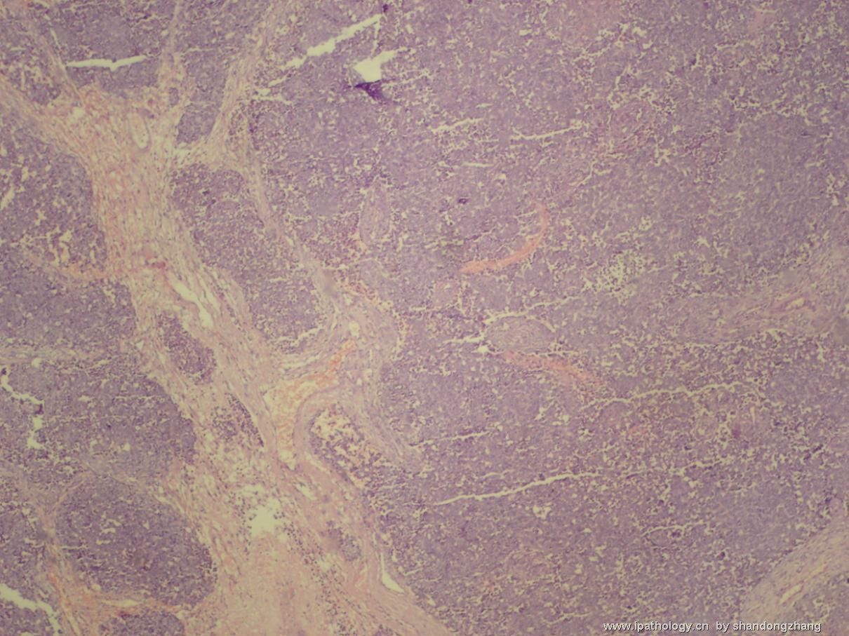
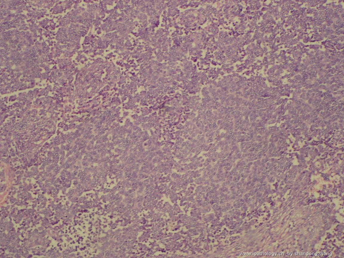
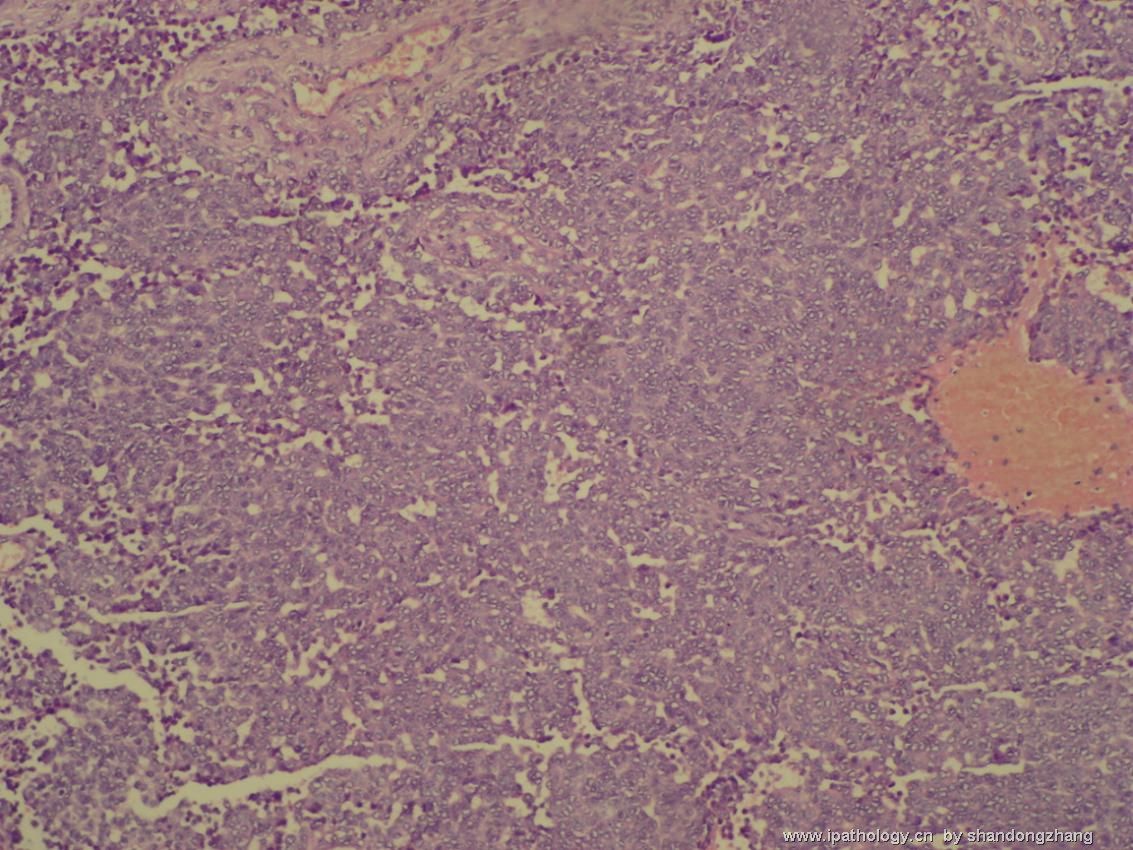
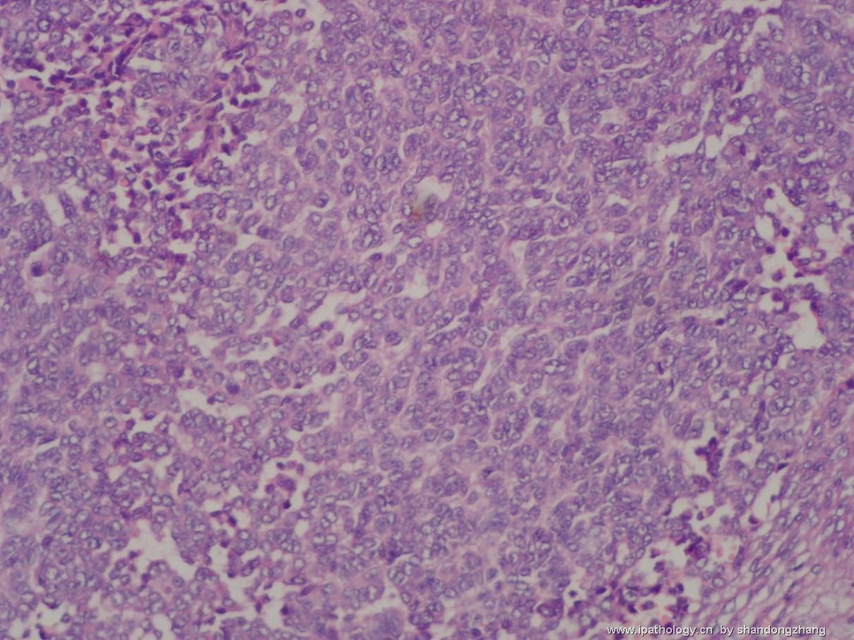
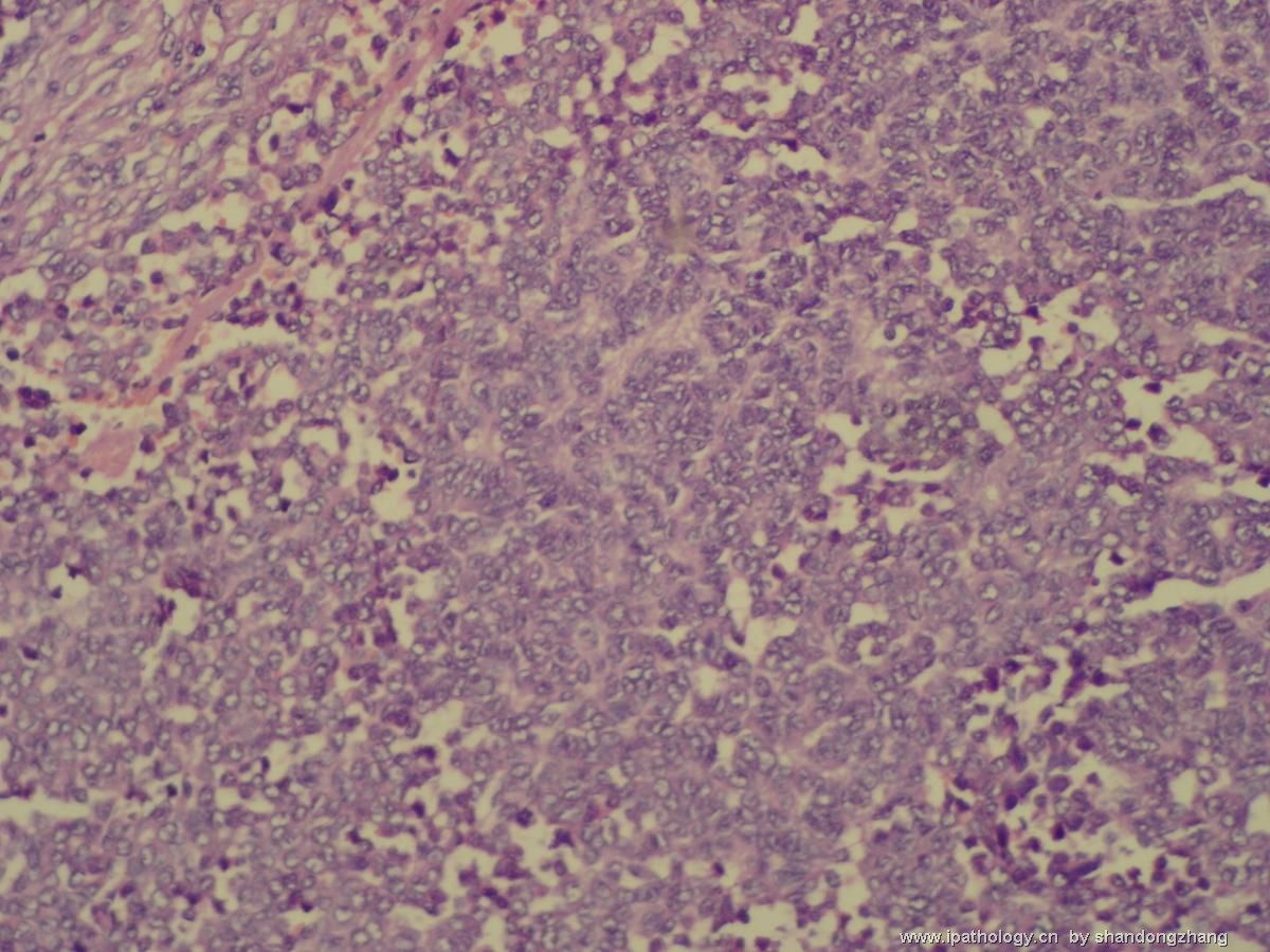
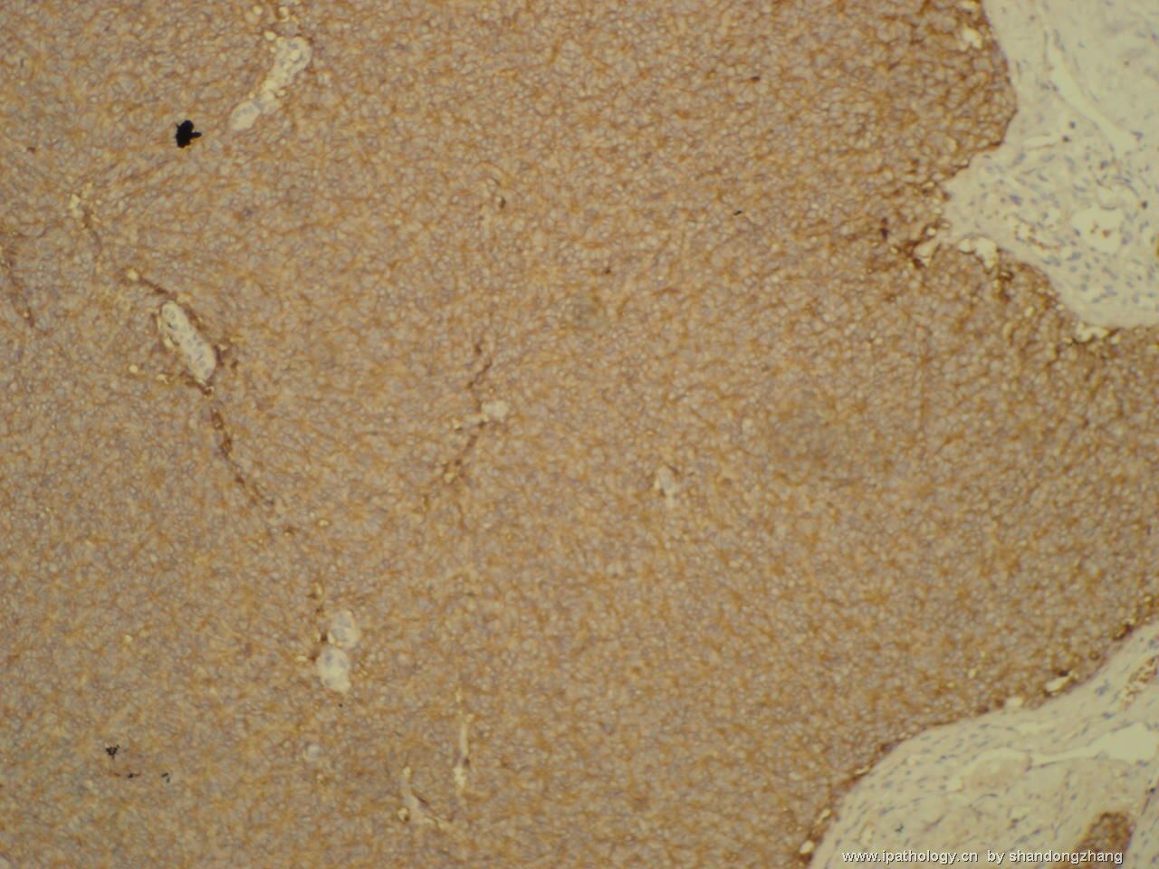
A cerebellar tumor like this, occurring in a 21 year old, generates a list of differential diagnosis that includes medulloblastoma (anaplastic or large cell variant), atyical rhabdoid/teratoid tumor (AT/RT), and less likely, choroid plexus carcinoma and metastasis. Distinction between these possibilities depends on clinical history, MRI finding and immunohistochemistry. Histologic features of anaplastic medulloblastoma overlap with that of AT/RT. AT/RT often contains mesenchymal areas of spindle cells, whic are not seen here. The last photo appears to be synaptophysin immunostain, which is strongly positive in neoplastic cells. If so, anaplastic/large cell medulloblastoma would be favored. This said, I would like to see more photos before making a final diagnosis.

聞道有先後,術業有專攻
-
Malignant meningioma or rhabdoid meningioma may have similar histopathology, but the immunohistochemical staining pattern shown is not consistent with any antibody commonly used for meningioma. The neoplastic cells do form Homer-Wright rosettes, which are again consistent with medulloblastoma of the large cell/anaplastic type. I would love to be corrected of my interpretation.

聞道有先後,術業有專攻
| 以下是引用mjma 在2006-10-7 13:37:00的发言: A cerebellar tumor like this, occurring in a 21 year old, generates a list of differential diagnosis that includes medulloblastoma (anaplastic or large cell variant), atyical rhabdoid/teratoid tumor (AT/RT), and less likely, choroid plexus carcinoma and metastasis. Distinction between these possibilities depends on clinical history, MRI finding and immunohistochemistry. Histologic features of anaplastic medulloblastoma overlap with that of AT/RT. AT/RT often contains mesenchymal areas of spindle cells, whic are not seen here. The last photo appears to be synaptophysin immunostain, which is strongly positive in neoplastic cells. If so, anaplastic/large cell medulloblastoma would be favored. This said, I would like to see more photos before making a final diagnosis. |
| 以下是引用mjma 在2006-10-10 2:36:00的发言: Malignant meningioma or rhabdoid meningioma may have similar histopathology, but the immunohistochemical staining pattern shown is not consistent with any antibody commonly used for meningioma. The neoplastic cells do form Homer-Wright rosettes, which are again consistent with medulloblastoma of the large cell/anaplastic type. I would love to be corrected of my interpretation. |
-
diandianfei 离线
- 帖子:35
- 粉蓝豆:12
- 经验:40
- 注册时间:2008-10-06
- 加关注 | 发消息

