| 图片: | |
|---|---|
| 名称: | |
| 描述: | |
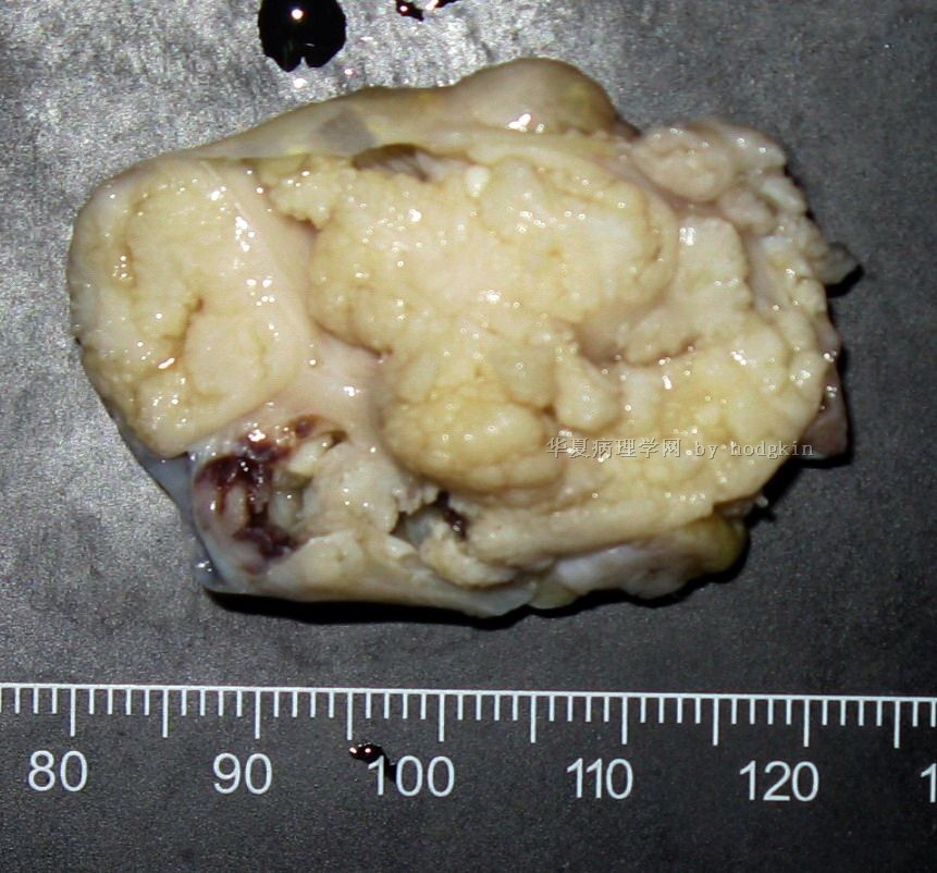
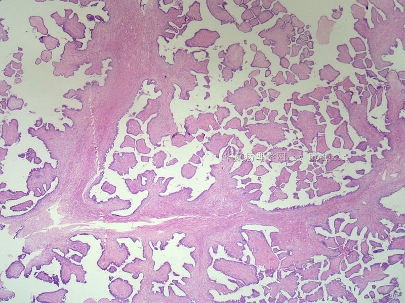
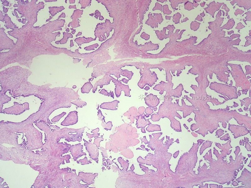
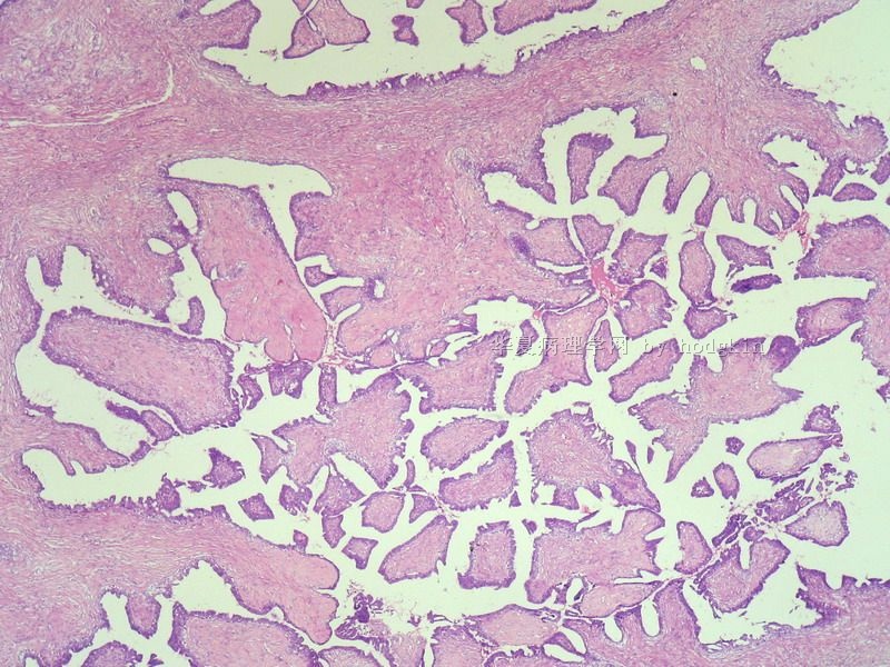
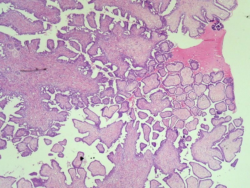
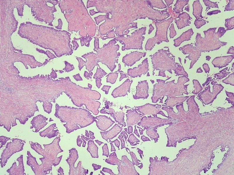
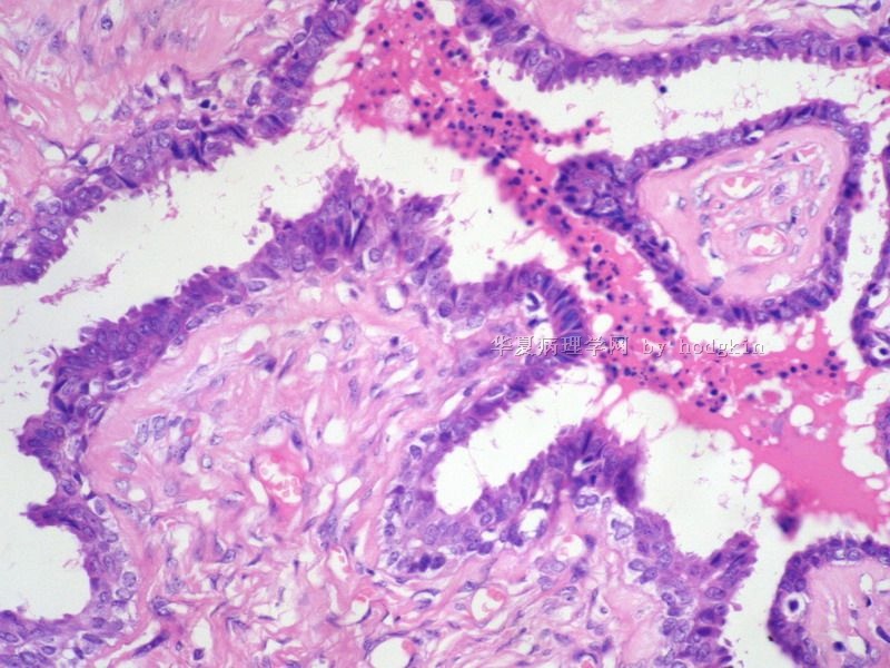
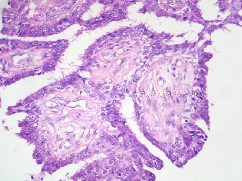
- B181816岁乳腺肿物。
-
本帖最后由 于 2009-07-13 22:48:00 编辑
Review the case and above interpretation carefully. The main differential dx includ 良性叶状肿瘤 and 糼年性纤维腺瘤.
Summary the features:
16 y/f-breast mass
Size: 4cm (based on the gross photo
Microscopically:
Cystic and papillary growth pattern
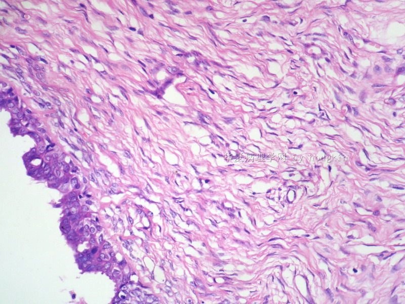
Stromal cellularity: mildly increased
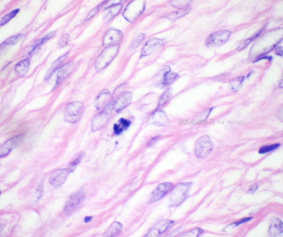
Stromal pleomorphism: mild to moderate (high power photo)
Stromal mitoses 0-1/10HPF
Stromal pattern uniform distribution and no heterologous elements
Margins: well-circumscribed.
I favor a diagnosis of cystic papillary benign phyllodes tumor as Dr. Chiang mentioned above.
Classic features of juvenile FA include increased stromal cellularity and epithelial hyperplasia. I cannot appreciate these in this case. Also mitoses are rarelly detected in the stroma in juvenile FA.
We always need to consider what clinicians will do when we have our diagnosis. The treatment will be the same for this case (complete excision) no matter what we call, benign phyllodes or juvenile FA.
Philosophy:
Mostly the young girl will be cured if she gets complete excision with clear margins. You will feel good for your dx (both FA and phyllodes) if the tumor has no recurrence in future. You will feel bad for your FA dx if the girl has local recurrence in the future.
仔细看了病例和以上讨论。主要鉴别诊断包括:叶状肿瘤和幼年性纤维腺瘤。
总结一下病变特征:
女性,16岁,乳腺肿块
大小:4cm(根据大体照片)
镜下:囊性,乳头状生长
间质细胞密度:轻度增加
间质细胞多形性:轻到中度(根据高倍图)
间质核分裂:0-1/10HPF
间质细胞分布均匀,没有异源性成分
边界:清楚
我倾向于诊断为囊性乳头状良性叶状肿瘤,与Dr. Chiang一致。
幼年性FA的典型特征包括间质细胞密度增加和上皮增生。本例我不能观察到这些。而且幼年性FA的间质细胞罕见核分裂。
我们作出诊断时总要考虑到临床后果。不管我们诊断为良性叶状肿瘤或幼年性FA,临床处理同样是完整切除。
经验总结:
如果完整切除,切缘干净,绝大多数年轻女性都会治愈。如果将来肿瘤不复发,你会对诊断满意(不管是诊断为FA或叶状肿瘤)。如果将来复发,你会为诊断为FA而不安。(abin译)

