| 图片: | |
|---|---|
| 名称: | |
| 描述: | |
- 欣赏一例
-
liguoxia71 离线
- 帖子:4174
- 粉蓝豆:3122
- 经验:4677
- 注册时间:2007-04-01
- 加关注 | 发消息
-
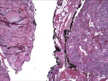
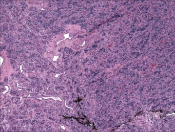
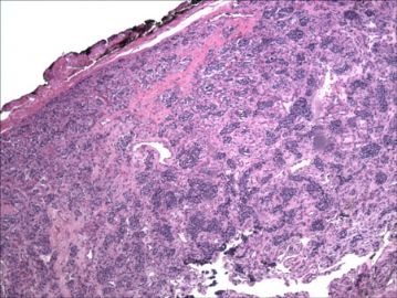
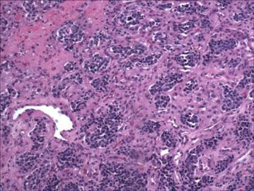
The tumor had a broad pushing front and is surrounded by a thin fibrous capsule. The tumor cells are arranged in a prominent nesting pattern, and the nests were separated by a delicate stroma with thin-walled vessels. The tumor cells had round to oval vesicular nuclei and occasional small nucleoli. Mitotic activity is low. There is no evidence of amyloid deposition or C-cell hyperplasia in the surrounding thyroid tissue.
肿瘤前缘呈广泛的推挤状,有薄层纤维包膜。肿瘤细胞明显巢状排列,细胞巢被含薄壁血管的纤细间质所分隔。细胞有圆形或椭圆形泡状核,偶见小核仁,核分裂少,周围甲状腺组织无淀粉样沉积或c细胞增生表现。

- jx16
-
Immunohistochemically, the tumor cells are strongly immunoreactive for chromogranin A. An S-100 immunohistochemical stain demonstrated strong staining in compressed spindled cells at the periphery of the cellular nests. The tumor cells are not immunoreactive for calcitonin, cytokeratin AE1:AE3, cytokeratin CAM 5.2, CEA, TTF-1 or thyroglobulin.
免疫组织化学:瘤细胞嗜铬粒蛋白A阳性,细胞巢外周扁平梭形细胞S-100强阳性,降钙素、AE1:AE3、CAM5.2、CEA、TTF-1以及甲状腺球蛋白均阴性。

- jx16
-
liguoxia71 离线
- 帖子:4174
- 粉蓝豆:3122
- 经验:4677
- 注册时间:2007-04-01
- 加关注 | 发消息


