| 图片: | |
|---|---|
| 名称: | |
| 描述: | |
- Kidney needle biopsy
-
Dr. QU, thank for sharing these interesting cases. Pothologists in general hospitals in China do not see these renal disease cases often. You may need to give an online lecture for some basic knowledge for renal diseases. Xiaohe can arrange the time in your convenance. Thanks, cz
-
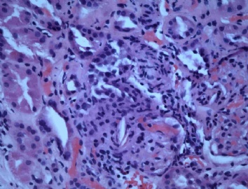
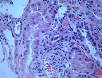
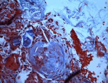
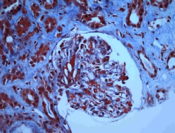
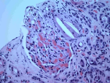
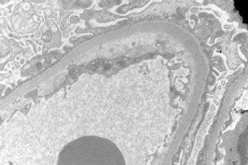
In this biopsy, the most striking change is the empty, needle-like clefts within the arterioles (photo 1, 2, 3, 5) and occasionally in capillaries (photo 4, 8). We may see multinucleated giant cell engulfing the clefts (photo 5). This is cholesterol embolism induced acute renal failure.
Photo 3 shows a globally sclerotic glomerulus with a cleft at the presumed arteriole (vascular pole). The cholesterol embolus is probably responsible for the death of that glomerulus. But it most likely occurred a while ago, not several weeks ago. I think this patient must have severe atherosclerosis at the aorta which ruptured and released the contents of atheroma to blood flow. The surgical event (endovascular grafting) made the situation worse, at least temporally.
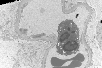
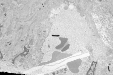
The last EM photo demonstrates subendothelial space widening. I interpret it as acute ischemic change. This type of change is also seen in thrombotic microangiopathy or transplant glomerulopathy. In this biopsy, it is focal and not severe enough. The thrombotic microangiopathy also does not fit the clinical picture.
-
本帖最后由 于 2009-06-24 17:58:00 编辑
我试着翻译一下:
这例活检最显著的改变是动脉中(图1,2,3,5),偶尔毛细血管中(图4,8)的空亮、针状裂隙 ,还可以看到多核巨细胞吞食裂隙现象(图5),这是引起急性肾衰的胆固醇栓子。
图3显示一个球状硬化的肾小球内包含一个可能位于动脉内的裂隙,这个胆固醇栓子可能是该小球死亡的原因。这个栓子更可能形成于不久前,而不是几周以前。病人主动脉一定有严重的动脉粥样硬化,随后粥样斑块破裂并将内容物释放进血流。 至少从时程上看,血管移植术(endovascular grafting)这一外科事件使情况变得更糟。
最后的电镜照片显示了内皮下空间增宽,我个人用急性缺血来做解释。这种改变也见于血栓形成的微血管和移植后肾病中。此例是局部的,并不严重。血栓形成的微血管也不符合这种临床图片。

- 嫁人就嫁灰太狼,学习要上华夏网。
|
有一点疑问的是:病人有急性肾功能衰竭,应该是绝大部分的肾小球都受累,这些胆固醇拴子来自那个血管呢?如果是来自主动脉等肾外的大血管,除了肾功能受损外,其它的脏器也应该受损,如脑、脾等,如果来自肾动脉,两个动脉同时发生胆固醇栓子的机会应该是比较少啊! These are good questions. Though this case is not my first one, I have not thought about these issues. I need do some reading and then answer these questions. |
| 以下是引用geng72在2009-6-18 10:34:00的发言:
很好的病例,第一次见到这样的病例。 有一点疑问的是:病人有急性肾功能衰竭,应该是绝大部分的肾小球都受累,这些胆固醇拴子来自那个血管呢?如果是来自主动脉等肾外的大血管,除了肾功能受损外,其它的脏器也应该受损,如脑、脾等,如果来自肾动脉,两个动脉同时发生胆固醇栓子的机会应该是比较少啊! |
Here is a recent reference which may answer the above questions:
Cholesterol crystal embolism: A recognizable cause of renal disease.
Division and Chair of Nephrology and Department and Chair of Pathology, Spedali Civili and University, Brescia, Italy. fscolar@tin.it
Cholesterol crystal embolism, sometimes separately designated atheroembolism, is an increasing and still underdiagnosed cause of renal dysfunction antemortem in elderly patients. Renal cholesterol crystal embolization, also known as atheroembolic renal disease, is caused by showers of cholesterol crystals from an atherosclerotic aorta that occlude small renal arteries. Although cholesterol crystal embolization can occur spontaneously, it is increasingly recognized as an iatrogenic complication from an invasive vascular procedure, such as manipulation of the aorta during angiography or vascular surgery, and after anticoagulant and fibrinolytic therapy. Cholesterol crystal embolism may give rise to different degrees of renal impairment. Some patients show only a moderate loss of renal function; in others, severe renal failure requiring dialysis ensues. An acute scenario with abrupt and sudden onset of renal failure may be observed. More frequently, a progressive loss of renal function occurs over weeks. A third clinical form of renal atheroemboli has been described, presenting as chronic, stable, and asymptomatic renal insufficiency. The renal outcome may be variable; some patients deteriorate or remain on dialysis, some improve, and some remain with chronic renal impairment. In addition to the kidneys, atheroembolization may involve the skin, gastrointestinal system, and central nervous system. Renal atheroembolic disease is a difficult and controversial diagnosis for the protean extrarenal manifestations of the disease. In the past, the diagnosis was often made postmortem. However, in the last decade, awareness of atheroembolic renal disease has improved, enabling us to make a correct premortem diagnosis in a number of patients. Correct diagnosis requires the clinician to be alert to the possibility. The typical patient is a white man aged older than 60 years with a baseline history of hypertension, smoking, and arterial disease. The presence of a classic triad characterized by a precipitating event, acute or subacute renal failure, and peripheral cholesterol crystal embolization strongly suggests the diagnosis. The confirmatory diagnosis can be made by means of biopsy of the target organs, including kidneys, skin, and the gastrointestinal system. Thus, Cinderella and her shoe now can be well matched during life. Patients with renal atheroemboli have a dismal outlook. A specific treatment is lacking. However, it is an important diagnosis to make because it may save the patient from inappropriate treatment. Finally, recent data suggest that an aggressive therapeutic approach with patient-tailored supportive measures may be associated with a favorable clinical outcome.
自学看了几天肾脏超微结构的书,还是觉得没能看明白。
第二次作业(学看本例超微结构):
超微图1:内皮细胞基膜局限性增厚,伴电子致密物沉积;小灶上皮细胞足突融合;毛细血管腔内分叶核细胞浸润?
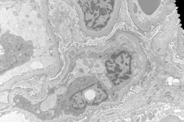
超微图2:似乎是系膜细胞插入内皮下?
超微图3:似乎是系膜基质增多,伴中等度的电子致密物沉积;左下方深染的是什么呢?似乎是个核的切缘;
超微图4:足突融合;内皮细胞下基膜内侧出现疏松化结构?
复习一下HE:图1似乎是肾小球血管极外血管病变?
图2:小血管壁增生腔狭窄?近端小管上皮颗粒状透明性改变是否为高蛋白尿所致?右侧肾小管上皮似乎有部分增生?
图4、图5似有系膜细胞增生。
题目全作答了,待我回头学习楼上各位专家、同道的回贴!谢谢!

- “人生没有彩排,每一天都是现场直播”
Hi, 197, Here is your decription of the above EM photo:
超微图1:内皮细胞基膜局限性增厚,伴电子致密物沉积;小灶上皮细胞足突融合;毛细血管腔内分叶核细胞浸润?
内皮细胞基膜局限性增厚
The glomerular basement membrane (GBM) is not obviously thickened. Some segments of GBM appear thickened probably due to tangential section. There are two ways to judge the thickness of the GBM. 1) Using the average width of the foot process: the "normal" thickness of GBM should roughly equal to the average width of the foot process. 2) Doing the actual measurement of the GBM: today more and more electron microscopes are equipped with digital cameras. It is easy to measure the thickness of GBM on the computer. The problem is how to define the normal range of GBM thickness. There is no standard range. Based on my reading, 250 nm could be used as lower limit and 460 nm could be considered as upper limit.
伴电子致密物沉积
There are no electron dense deposits.
小灶上皮细胞足突融合
There is little foot process fusion or effacement. We could ignore it in this photo.
毛细血管腔内分叶核细胞浸润?
Yes, you are right!
-
本帖最后由 于 2009-06-30 20:39:00 编辑
似乎是系膜基质增多,伴中等度的电子致密物沉积;左下方深染的是什么呢?似乎是个核的切缘;
These photos were taken by a technician. I am not entirely sure that the capillary in this photo is (intra)glomerular capillary or interstitial capillary. I guess it is a capillary or arteriole close to glomerular vascular pole. Therefore, there is no well-defined mesangium. No electron dense deposits are identified, either. 左下方深染的是grid bar. The plastic sections are put on the grid to be examined under electron microscope. The grid is made of copper, Ni or gold.
非常感谢quhong老师百忙中答疑解惑!!!


特别邀请fxzxyy作为我的网络同桌一起认真学习quhong老师的解答!
特别特别邀请wfbjwt得空时友情客串华夏病理学网quhong老师的翻译官!注:别担心没时间,首先时间象海绵,呵呵;其次,我们总是在您得空时才邀请,呵呵;第三别担心把网友们宠坏了,至少——
还要邀请英语不太好的网友们和我一起学习读英文,自己心头翻译一番再与wfbjwt老师的答案来对照,呵呵。注:别担心他的不是标准答案,因为,万一他翻译错了,楼主老师一定会帮他。

- “人生没有彩排,每一天都是现场直播”
