


| 图片: | |
|---|---|
| 名称: | |
| 描述: | |
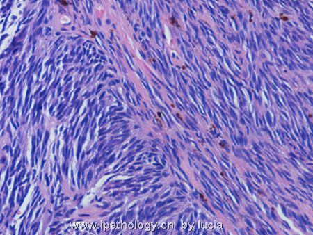
- 颅内肿瘤-转移性恶黑
Thank Dr Mjma very much for the differential diagnosis.
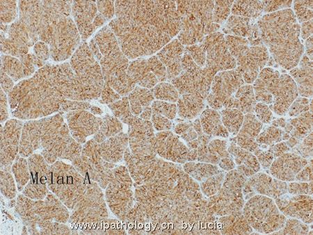
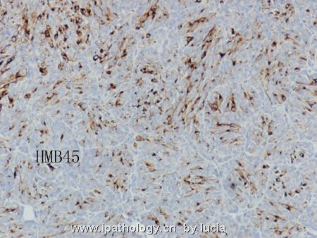
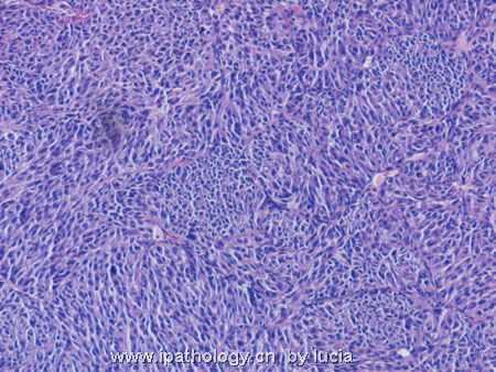
The diagnosis is metastatic melanoma.
This tumour demonstrated two different morphologies. In most areas the tumour cells are oval and arranged in nests separated by thin fibrovascular tissue. In other areas the tumour cells show elongated nuclei and arranged in fascicles (not shown last time). With these morphologies, both primary, ie gliosarcoma and malignant meningioma, and metastatic tumour have to be considered. With no identifiable fibrillary background in the entire tumour, glial tumour can be readily ruled out. Because of entirely intracerebellar growth, meningeal origin is also unlikely. Metastatic tumours should consider melanoma (coexists of both nested and spindled areas), neuroendocrine tumour (nests separated by fibrovascular septa) and poorly differentiated carcinoma. Immunohistochemistry shows that the tumour is positive for S-100, Melan-A and HMB45. However, no melanin pigments are identified. It is negative for cytokeratin, synaptophysin, chromogranin and GFAP.
The patient presented with no history of any primary lesion. She died soon after operation. The cranial lesion is solitary.
名称:图1
描述:图1
名称:图2
描述:图2
名称:图3
描述:图3
名称:图4
描述:图4


