| 图片: | |
|---|---|
| 名称: | |
| 描述: | |
- 顶枕部占位
-
liguoxia71 离线
- 帖子:4174
- 粉蓝豆:3122
- 经验:4677
- 注册时间:2007-04-01
- 加关注 | 发消息
-
本帖最后由 于 2009-06-07 10:51:00 编辑
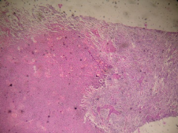
There are large cells with plump cytoplasm, vesicular nuclei and prominent nucleoli in a background of less well differentiated cells with oval or slightly elongated cells. Necrosis is seen. I suspect this is a malignant neoplasm with neuronal differentiation. How often do you find mitotic figures in the smaller cells? I recommend staining for synaptophysin, NSE, GFAP and MIB-1 to help us interpret this lesion.
liguoxia71试译,请大家指正:伴有卵圆或短梭细胞的分化程度不高的细胞背景中见一些胞浆丰富、核空泡状有突出核仁的大细胞,可见坏死,我怀疑是有神经元分化的恶性肿瘤。不知道小一些的细胞核分裂象多少。建议染syn、NSE、GFAP、MIB-1帮助我们诊断。

聞道有先後,術業有專攻
-
jixiangrui 离线
- 帖子:17
- 粉蓝豆:1
- 经验:17
- 注册时间:2009-05-18
- 加关注 | 发消息


