| 图片: | |
|---|---|
| 名称: | |
| 描述: | |
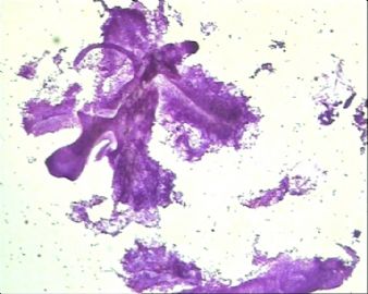
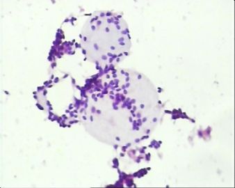
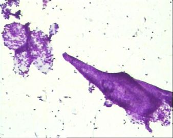
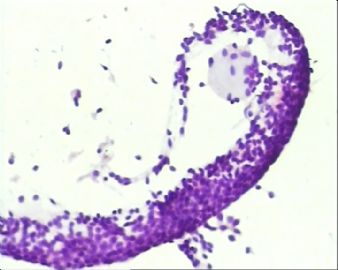
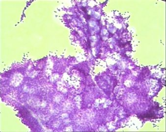
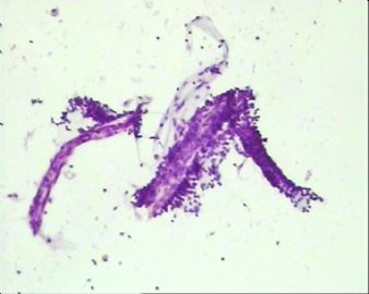
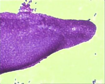
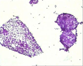
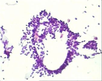
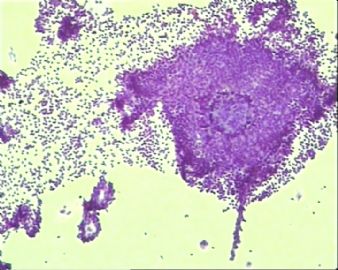
- 左上腭肿块穿刺,请诊断。
-
taotaochan 离线
- 帖子:131
- 粉蓝豆:22
- 经验:131
- 注册时间:2007-03-17
- 加关注 | 发消息
黏液球团状细胞 can be seen in the following salivary gland neoplasms: 1)Pleomorphic adenoma; 2) Basal cell adenoma; 3) Adenoid cystic carcinoma; 4) Myoepithelioma or carcinoma; 5) Polymorphous low-grade adenocarcinoma. So, it is not that specific, you have to look at the whole slide and the cells carefully, before you render the diagnosis.
I am trying to post a nice paper addressing this issue.oops! I can only post the abstract and not the pdf file of the whole paper. Maybe somebody can help me?
Cytologic diagnosis of adenoid cystic carcinoma of salivary glands.
Nagel H, Hotze HJ, Laskawi R, Chilla R, Droese M.
Department of Cytopathology, Georg-August-University of Göttingen, Germany. hnagel@med.uni-goettingen.de
The cytomorphologic features in fine-needle aspiration (FNA) biopsies from 31 primary and 33 recurrent adenoid cystic carcinomas (ACC) were investigated. The correct FNA diagnosis was established in 24 of 31 primary ACC (77%). The diagnostic clue in aspirates from ACC are large globules of extracellular matrix, partially surrounded by basaloid tumor cells. In FNAs with predominance of basaloid tumor cells, but lacking characteristic globules, all other benign and malignant salivary gland tumors of epithelial-myoepithelial differentiation should be considered in the cytologic diagnosis. Pleomorphic adenoma is most frequently confused with ACC, and therefore, the cytologic findings in FNAs from 50 pleomorphic adenomas were compared with those diagnosed as ACC. Furthermore, rare neoplasms of salivary glands with epithelial-myoepithelial cell differentiation, including basal-cell adenoma and carcinoma, epithelial-myoepithelial carcinoma, and polymorphous low-grade adenocarcinoma, as well as some nonsalivary gland neoplasms presenting an adenoid cystic pattern, must be considered. The cytologic features of these entities are discussed in detail with respect to the cytologic diagnostic criteria of ACC.
-
xianyuanqq82 离线
- 帖子:455
- 粉蓝豆:55
- 经验:456
- 注册时间:2009-02-04
- 加关注 | 发消息
This is a beautiful aspirate and I basically agree with most of the posts that it most likely represent an adenoid cystic carcinoma.
What size of the needle you used to do the aspiration? few photos really show a microbiopsy-like picture. I have one more question for those who are interested in FNA, can you see 黏液球团状细胞 in other salivary gland neoplam?

