| 图片: | |
|---|---|
| 名称: | |
| 描述: | |
- B1803少见乳腺肿物,鉴别诊断? (cqz-18, 5-7-2009)
| 姓 名: | ××× | 性别: | 年龄: | ||
| 标本名称: | |||||
| 简要病史: | |||||
| 肉眼检查: | |||||
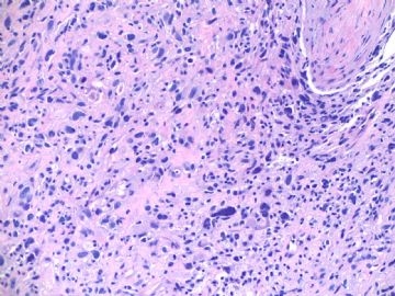
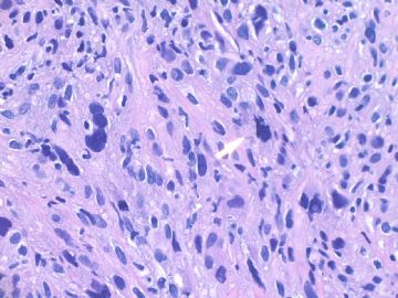
about 60 y/f, right breast mass 2 cm by imaging, breast core biopsy.
1. Your differential diagnosis?
2. What will you do next?
Please do not just give a diagnosis. Your dx is a guess dx even though your diagnosis may be right.
As pathologists we should learn how to analyse our cases with logic thought.We should have differential dx for all cases in our mind even for the easy cases. We can use available sources (such as IHC, molecular methods, history, consultation) to rule in or rule out the differential dx, then make the final dx.
Learning the priniciple for diagnosis is much more important than learning a few cases.
-
本帖最后由 于 2009-05-07 18:28:00 编辑
相关帖子
如果我遇到这样的病例,我的第一印象是:梭形细胞化生性癌---可以用CK7、CK8/18等确定其上皮性质;周围可能有原位癌成分;另外,梭形细胞癌也常标记肌上皮标记物(如P63、S-100、calponin等故需注意与肌上皮癌鉴别)。
当然一定要有其他鉴别诊断:1、恶性叶状肿瘤---寻找良性上皮成分;梭形细胞CK阴性而Vimentin阳性;或可有异源性分化(如骨、肌源性分化等)。2、乳腺间质肉瘤。3、乳腺非特异性软组织肉瘤。……
期待Dr.zhao的结果与分析。

- 博学之,审问之,慎思之,明辨之,笃行之。
-
lfl001200546 离线
- 帖子:2808
- 粉蓝豆:40
- 经验:2808
- 注册时间:2007-02-14
- 加关注 | 发消息
-
本帖最后由 于 2009-05-10 21:39:00 编辑
笃(du)行者 :“反复看,查文献,多取材,做特免,实在不行请外援”。Very good. I think I will not forget the word "笃".
This case was a breast core biopsy, only one tissue block. The photo represents the lesion in the available specimen.
More IHC results: ER, PR, Her2 negative.
Will you sing out the case (?diagnosis) or do more study? If you want to work more on this case, what will you do? What immunostains will you order? Why? You can order the immunostains. I am your lab technologists and do the stains for you and tell your the stain results.
I am in Los Angeles to enjoy my vocation time. Glad to work for this case with your guys.
cz
-
本帖最后由 于 2009-05-10 21:50:00 编辑
老年,乳腺的梭形细胞恶性肿瘤
鉴别诊断包括:上皮性--梭形细胞癌,肌上皮性--肌上皮癌,乳腺间质--叶状肉瘤,软组织肉瘤,其它(恶黑,少见淋巴造血肿瘤)。
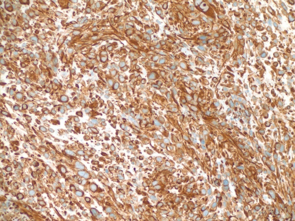
已知AE1/AE3和Cam 5.2均阴性,Vimentin弥漫强阳性,排除上皮性。进一步分类:
叶状肿瘤:ER,PR,
肌上皮:SMA,CP,p63
恶黑:S-100,HMB45,Melan
神经源:S-100,Syn,Cga
脂肪肉瘤:S-100
肌源性:SMA,Desmin,MyoD1,
血管:CD34,CD31
纤维母/肌纤母:CD34
其它:纤维肉瘤,……
也可以等待手术标本再诊断。

华夏病理/粉蓝医疗
为基层医院病理科提供全面解决方案,
努力让人人享有便捷准确可靠的病理诊断服务。
| 以下是引用abin在2009-5-10 21:46:00的发言:
老年,乳腺的梭形细胞恶性肿瘤 鉴别诊断包括:上皮性--梭形细胞癌,肌上皮性--肌上皮癌,乳腺间质--叶状肉瘤,软组织肉瘤,其它(恶黑,少见淋巴造血肿瘤)。 已知AE1/AE3和Cam 5.2均阴性,Vimentin弥漫强阳性,排除上皮性。进一步分类: 叶状肿瘤:ER,PR, 肌上皮:SMA,CP,p63 恶黑:S-100,HMB45,Melan 神经源:S-100,Syn,Cga 脂肪肉瘤:S-100 肌源性:SMA,Desmin,MyoD1, 血管:CD34,CD31 纤维母/肌纤母:CD34 其它:纤维肉瘤,…… 也可以等待手术标本再诊断。
Very good analyses with differential dx. Two things I do not agree: 1. 已知AE1/AE3和Cam 5.2均阴性,Vimentin弥漫强阳性,排除上皮性。Mostly you are right. But you may be wrong. Tell us why. 2.也可以等待手术标本再诊断. Completely do not agree.
|
to Dr. Zhao:
1. 已知AE1/AE3和Cam 5.2均阴性,Vimentin弥漫强阳性,排除上皮性。Mostly you are right. But you may be wrong. Tell us why.

我这样说是太绝对了。应该说:“AE1/AE3和Cam 5.2均阴性,Vimentin弥漫强阳性,仍不能完全排除上皮性。”
多形性癌:均表达AE1/AE3和Cam 5.2。
腺癌伴梭形细胞化生:表达CK7。
化生性癌或癌肉瘤:其中的异源性化生的梭形细胞成分,可以局灶性表达CK,当然也可能不表达。可以加做CK7和34BE12。
2.也可以等待手术标本再诊断.
Completely do not agree.
这涉及到穿刺标本取材的局限性,是否只取到梭形细胞成分而不见典型的浸润癌成分。正因为如此,我觉得穿刺标本诊断困难时,先报“恶性肿瘤”,等待手术标本再诊断,也许可以避免困难和风险。
不知我的理解是否正确?期盼Dr.Zhao进一步指导。谢谢!

华夏病理/粉蓝医疗
为基层医院病理科提供全面解决方案,
努力让人人享有便捷准确可靠的病理诊断服务。
-
本帖最后由 于 2009-05-15 19:42:00 编辑
From Abin
这涉及到穿刺标本取材的局限性,是否只取到梭形细胞成分而不见典型的浸润癌成分。正因为如此,我觉得穿刺标本诊断困难时,先报“恶性肿瘤”,等待手术标本再诊断,也许可以避免困难和风险。
You are right in some points.
We should try to provide clinicians and patients as more information as we can from the biopsy specimens. It is perfect if we can have final diagnosis in the biopsy specimen. Clinicians can decide next procedure based on pathology diagnosis from biopsy. The treatment plans may be different if this patient has metastatic lesion, melaloma, carcinoma or sarcoma. Of cause we may not make the definite dx sometimes based on the biopsy specimen after we try.
你的部分观点的正确的。
对于活检标本,我们应该努力为临床医生和患者提供尽可能多的信息。如果活检标本能作出最终诊断,它是很完美的。临床医生可以根据活检诊断决定进一步处理程序。如果是转移性病变、恶黑、癌或肉瘤,临床处理方案可能不同。当然,我们有时也可能在尽力之后仍不能根据活检标本作出明确诊断。(abin译)
-
本帖最后由 于 2009-05-15 19:45:00 编辑
In fact it is not very difficult to have the differential dx for this case.
1. The main ddx include sarcoma, carcinoma and others (melanoma, hematologic lesions et al).
2. It is metaplastic carcinoma or basal-like carcinoma if it is carcinoma.
3. What type of sarcoma if it is sarcoma? or just part of malignant phyllodes?
实际上本例的鉴别诊断并不太难。
1.主要鉴别:癌、肉瘤和其它(恶黑、淋巴造血系统病变等)。
2.如果是癌:转移癌或基底样癌。
3.如果是肉瘤:什么类型?或仅仅是恶性叶状肿瘤的一部分?(abin译)
-
本帖最后由 于 2009-05-15 19:47:00 编辑
If we want to rule out metaplastic carcinoma we must do more cytokeratin markers. Negative reaction to AE1/AE3, cam 5.2 is not enough.
More IHC I did for this case:
Negative p63, ck7, ck20, ck14, ck17, ck5/6, 34b-E12 (high molecular cytokeratin), EMA.
Negative for S-100, CD45 (LCA)
如果需要排除转移性癌,我们必须做更多上皮性标记物。AE1/AE3和cam 5.2阴性还不够。
我做了更多免疫组化如下:
阴性:p63, ck7, ck20, ck14, ck17, ck5/6, 34b-E12 (high molecular cytokeratin), EMA, S-100, CD45 (LCA)


