| 图片: | |
|---|---|
| 名称: | |
| 描述: | |
- 宫内膜
| 姓 名: | ××× | 性别: | 女 | 年龄: | 57 |
| 标本名称: | 宫内膜 | ||||
| 简要病史: | 停经5年,不规则阴道流血10天,超声示内膜厚9毫米 | ||||
| 肉眼检查: | |||||
名称:图1
描述:图1
名称:图2
描述:图2
名称:图3
描述:图3
名称:图4
描述:图4
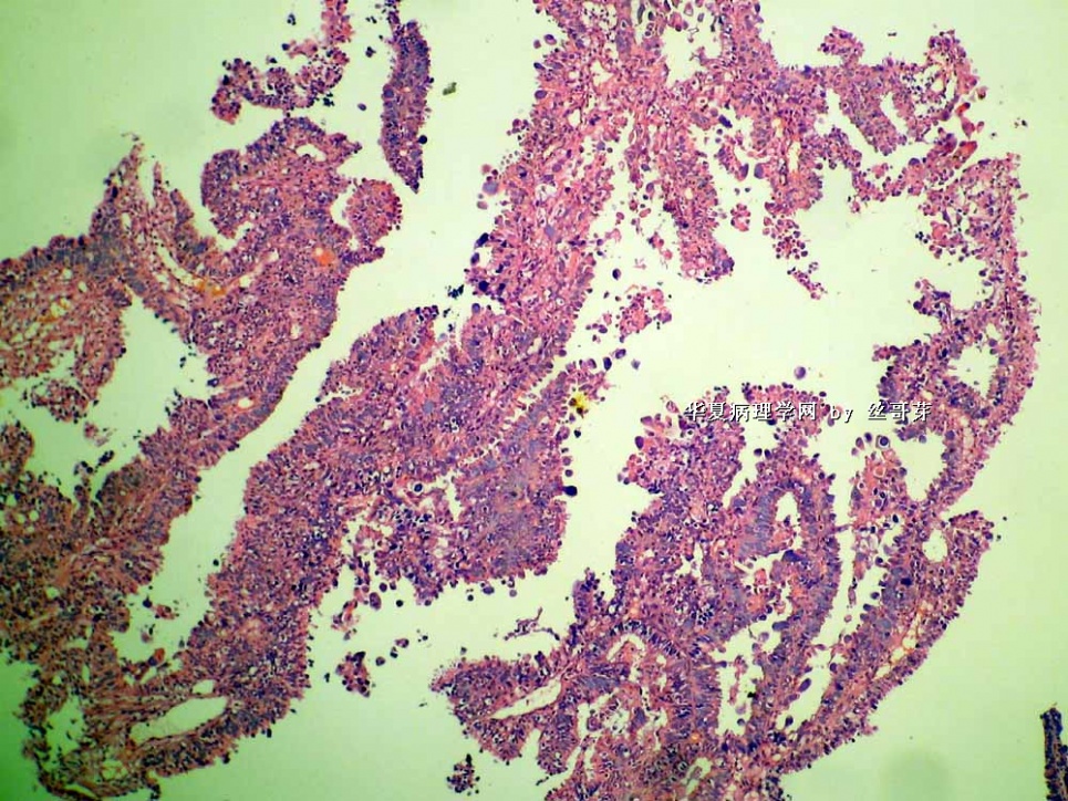
名称:图5
描述:图5
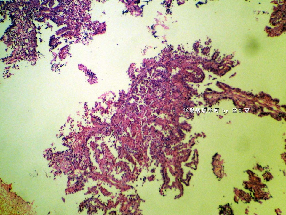
名称:图6
描述:图6
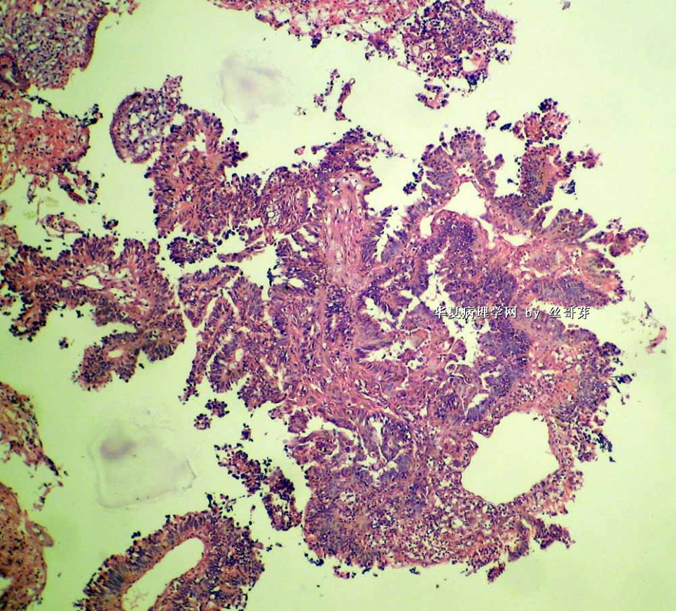
名称:图7
描述:图7
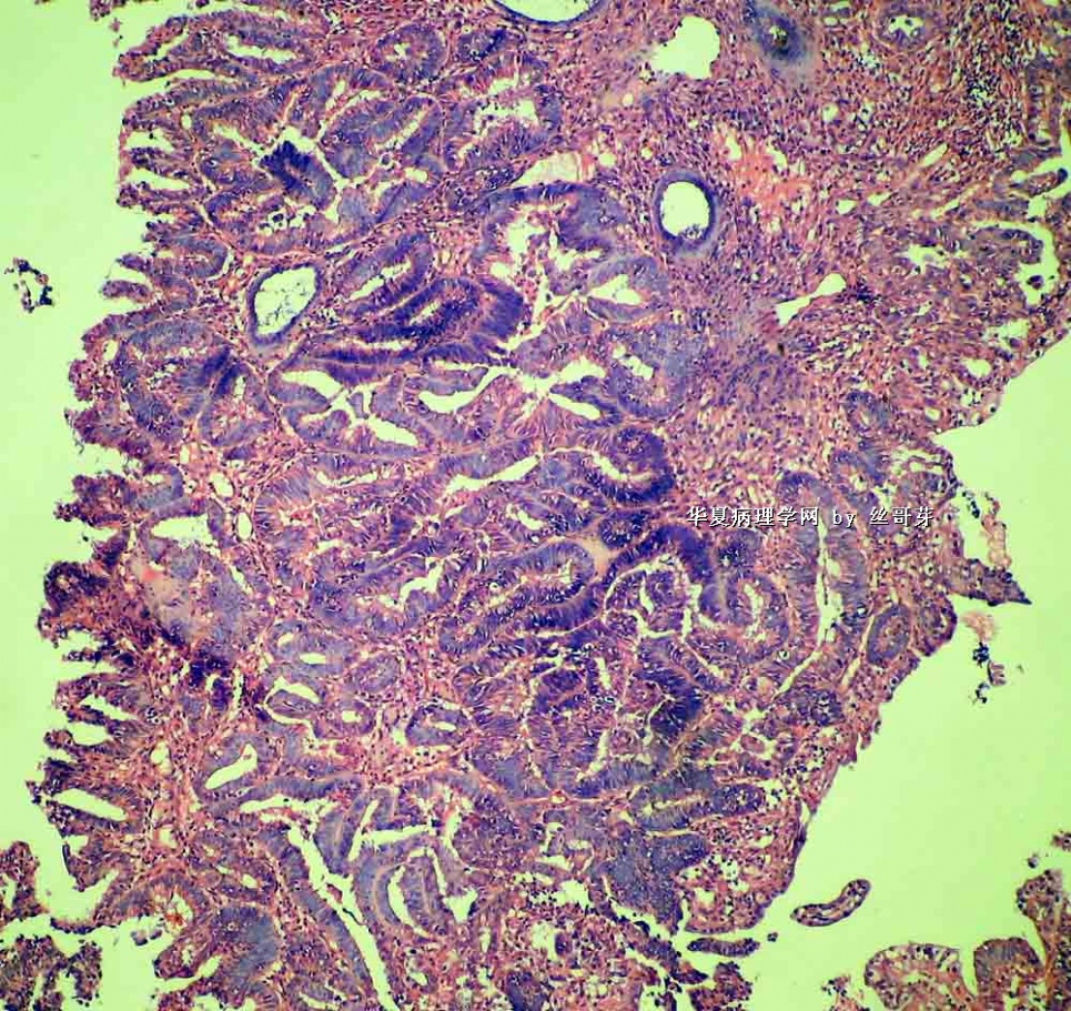
名称:图8
描述:图8
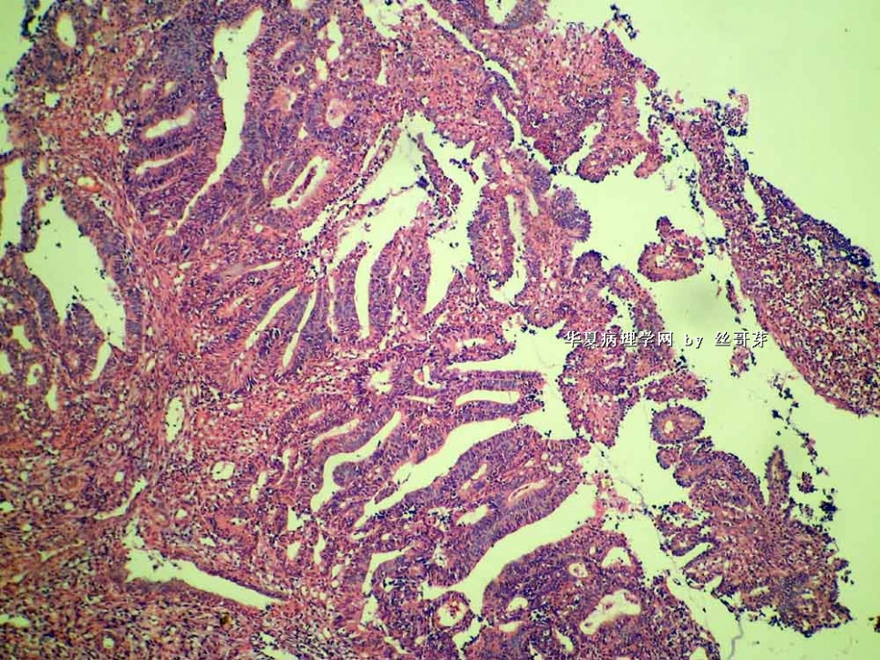
名称:图9
描述:图9
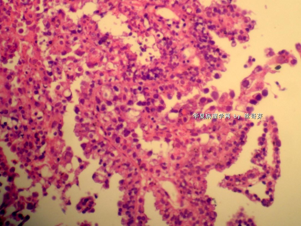
名称:图10
描述:图10
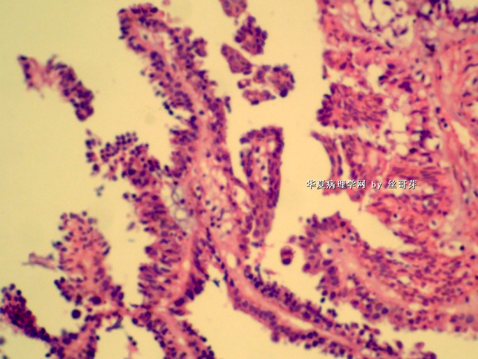
名称:图11
描述:图11
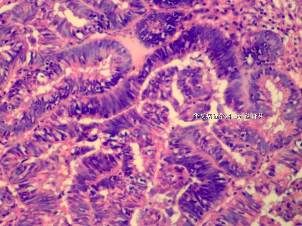
名称:图12
描述:图12
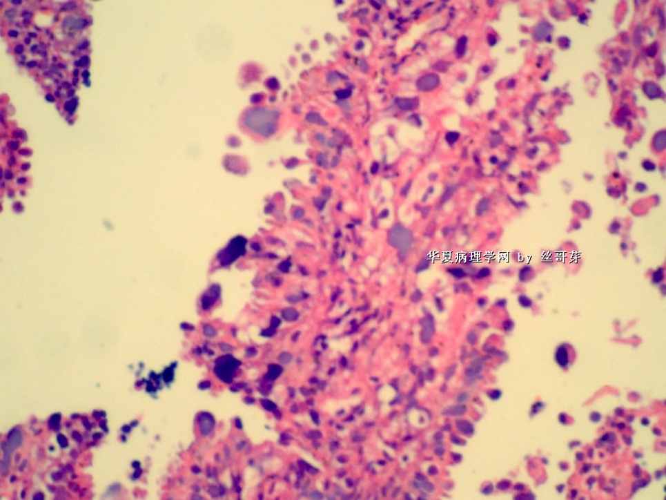
名称:图13
描述:图13
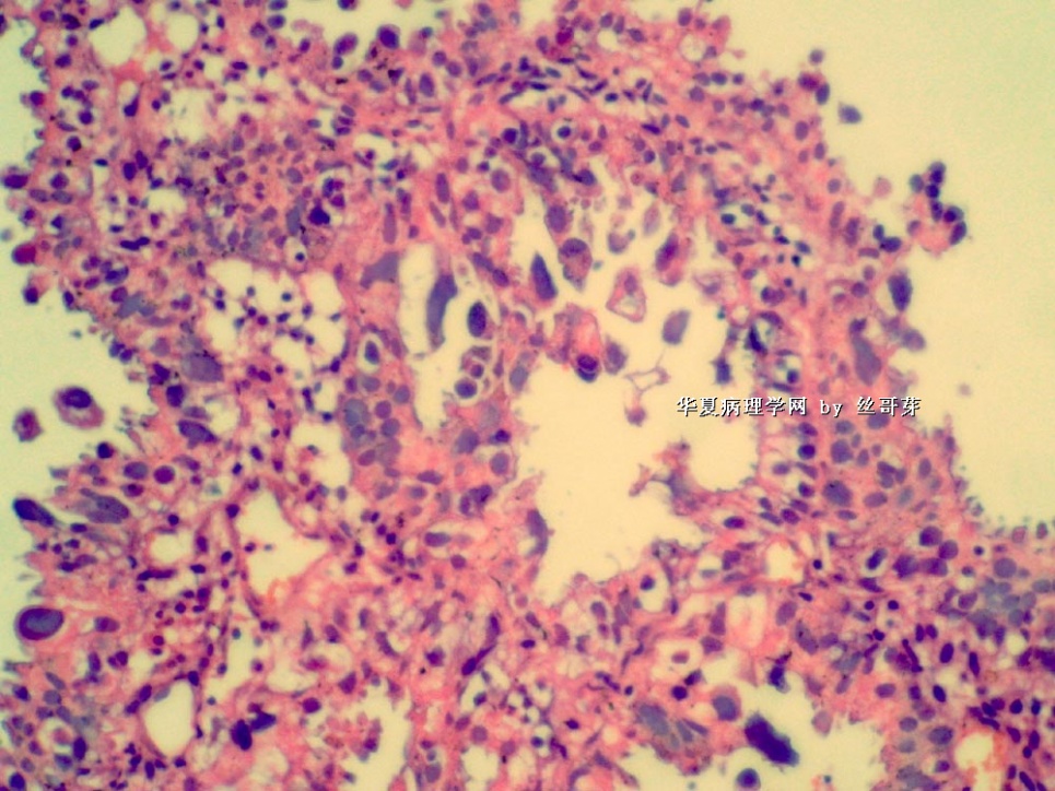
名称:图14
描述:图14
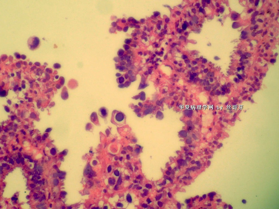
名称:图15
描述:图15
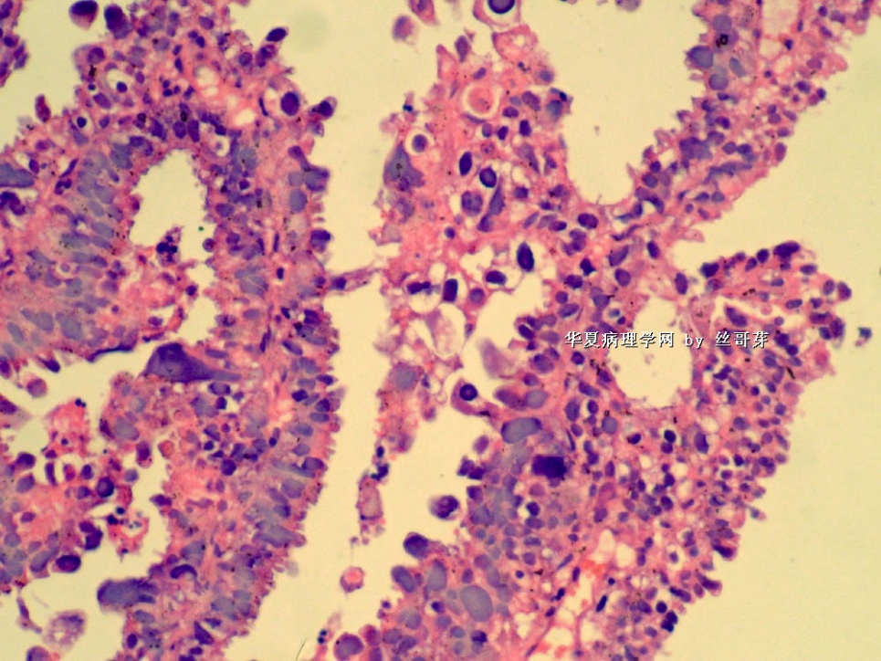
名称:图16
描述:图16
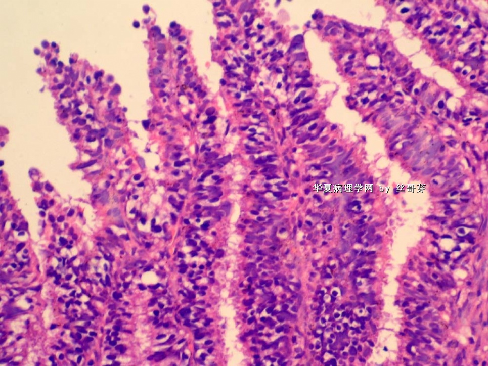
名称:图17
描述:图17
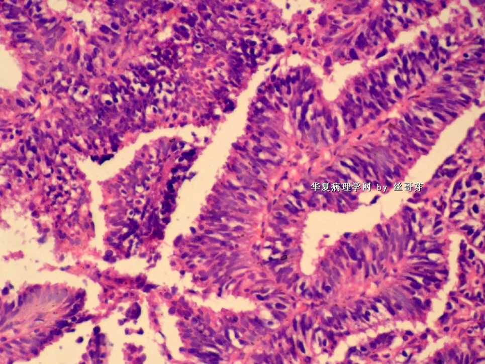
名称:图18
描述:图18
-
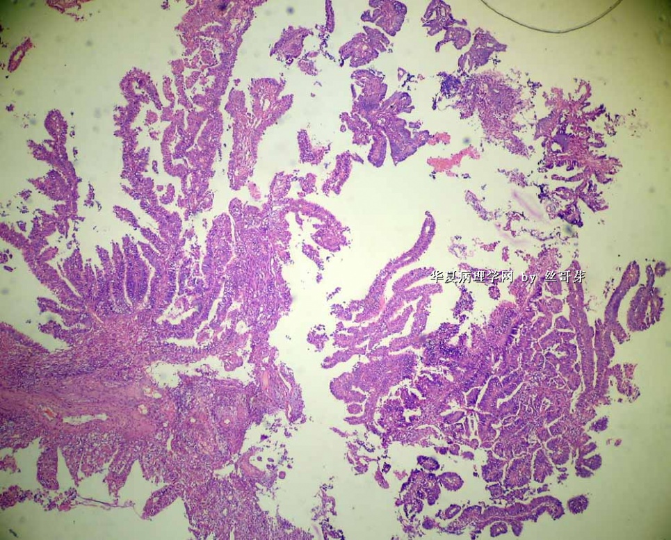
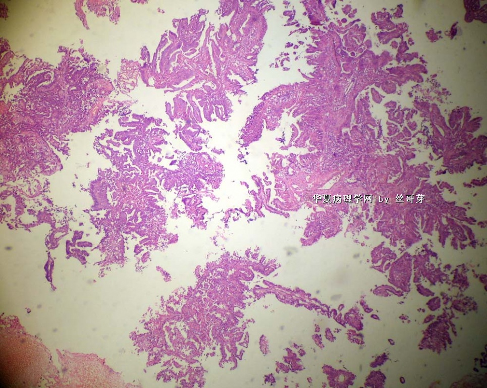
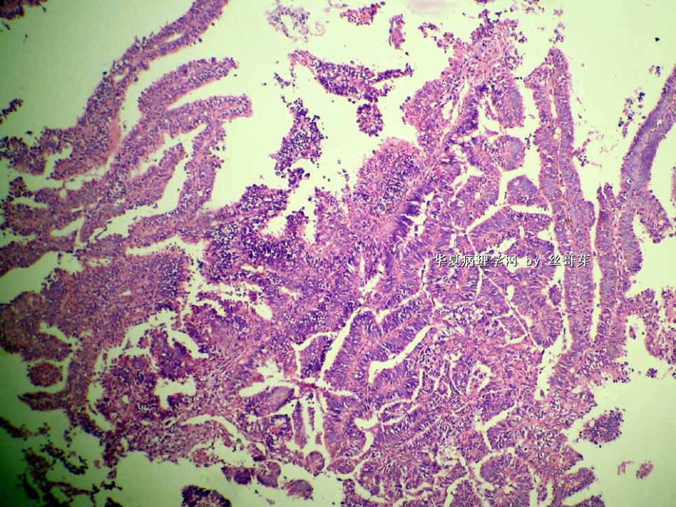
这可能是低分化内膜样腺癌,乳头状的
不太像浆液性的(UPSC)。不过也可能是,要免疫组化才能确定
为什么说不像?
UPSC:核一般是圆形,而不是柱状
细胞没有垂直于基底膜排列的极向
核常位于顶部而不是基底
有大量脱落的细胞团,而不是真的小乳头
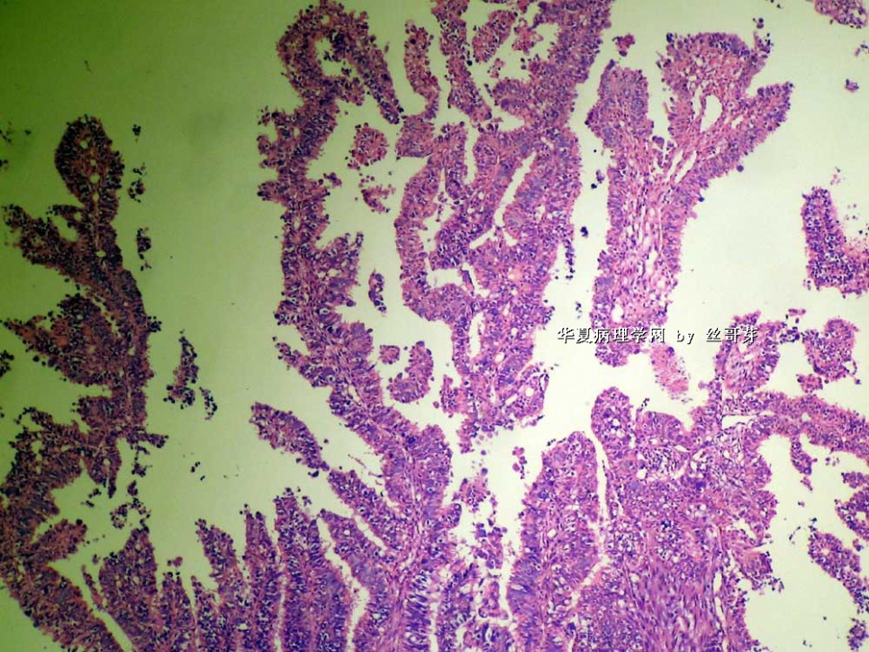
核异型性非常明显,有大的嗜酸性核仁
有的图上全是垂直排列的柱状细胞
也有的地方像,核异型性非常大,但是它们没有明显的核仁
那么,它有几种可能:
高级别的子宫内膜样癌,可能伴有假象
混合性(内膜样+UPSC)
纯UPSC的可能性很小
不过最终要看免疫组化,UPSC:ER- PR- P53+ Ki67高
如果不确定,报低分化腺癌

华夏病理/粉蓝医疗
为基层医院病理科提供全面解决方案,
努力让人人享有便捷准确可靠的病理诊断服务。
-
diandianfei 离线
- 帖子:35
- 粉蓝豆:12
- 经验:40
- 注册时间:2008-10-06
- 加关注 | 发消息
-
本帖最后由 于 2009-05-07 23:13:00 编辑
| 以下是引用abin在2009-5-4 21:53:00的发言: 这可能是低分化内膜样腺癌,乳头状的 |
Abin, good analysis and discussion! I basically agree with abin here. It looks like a typical "villoglandular endometrioid adenocarcinoma".
Couple photos at the end show "clearing changes" in the cytoplasm. I am not sure they represent the real "clear cell carcinoma" or secretory changes. I would like to see the low power picture in those clearing areas. My hunch is that they are most likely secretory changes, rather than true components of mixed clearing cell carcinoma. If on your slides you have any doubts, I will advise to go back your specimen and submit more sections to make sure you do not have clear cell carcinoma components, since the latter will disctate the clinical prognosis.
abin译:基本同意abin分析。后两图胞浆有“透明改变”。我不确定它代表真性的“透明细胞癌”或分泌改变。我想看透明区的低倍。我估计更可能像分泌改变,而不是真性混合有透明细胞癌。如果对切片上的病变有任何疑问,我建议再检查标本,补充取材,以确信没有透明细胞癌成分,因为后者预后差。

- 不坠青云之志,长怀赤子之心
-
wangxuanju 离线
- 帖子:113
- 粉蓝豆:1
- 经验:113
- 注册时间:2009-04-12
- 加关注 | 发消息
-
本帖最后由 于 2009-05-07 23:03:00 编辑
绒毛腺管状腺癌:是子宫内膜样腺癌的一个亚型。
在30%的内膜样腺癌中,有某种程度的绒毛腺样结构。然而,以这种类型为主的子宫内膜样癌不足5%(Crum CP)。
绒毛腺管状腺癌是以出现细长乳头为特征的高分化肿瘤,可能主要是典型的子宫内膜样或完全为乳头状癌。细胞学特征与典型的子宫内膜癌一样,细胞核通常具有1级核的特征。这些肿瘤分化好,因此,通常有较好的预后。然而,具有浸润成分的绒毛腺管乳头状结构的肿瘤可能伴有较差的预后(Anderson MC,et al)。
本例,出现高级别核,似乎不应再归入绒毛腺管状腺癌,也不应认为是高分化子宫内膜样腺癌。当然,最终诊断要看手术标本中的病变。
