| 图片: | |
|---|---|
| 名称: | |
| 描述: | |
- Neck FNA
-
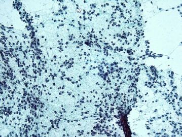
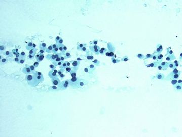
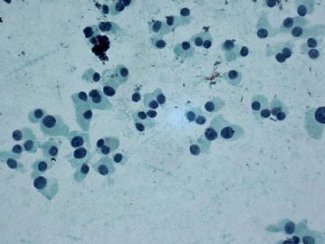
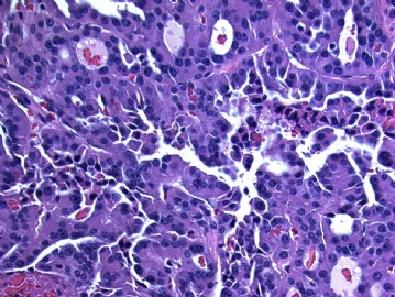
Just got this case early this week. This is a 60-year-old female had a total thyroidectomy in 2004 for papillary thyroid carcinoma. Now she has a 1.5 cm nodule developed in the neck. What's your diagnosis/differential diagnosis?
-
liguoxia71 离线
- 帖子:4174
- 粉蓝豆:3122
- 经验:4677
- 注册时间:2007-04-01
- 加关注 | 发消息
-
Ok. I did some immunostains on the cell block, the cells are negative for calcitonin and CEA. I would like to try more stains, but only got limited amount of cells on cell block. The thyroid surgeon also checked her serum calcitonin, which is normal. My question to this forum is: can any papillary thyroid carcinoma look like this? Before you give the patient second neoplasm, you better make sure that this is not recurrent papillary carcinoma, which is more likely to the clinicians?
-
Ok. I did some immunostains on the cell block, the cells are negative for calcitonin and CEA. 好的,我在细胞块上做了一些免疫组化,降钙素和CEA都是阴性的,I would like to try more stains, but only got limited amount of cells on cell block. 我还想做更多的免疫组化,但是因细胞块太少,做不了那么多免疫组化。The thyroid surgeon also checked her serum calcitonin, which is normal. 甲状腺手术医生也做了血清学的降钙素也是正常的。My question to this forum is: can any papillary thyroid carcinoma look like this? 我问题是这个图象能说是乳头状癌吗?Before you give the patient second neoplasm, you better make sure that this is not recurrent papillary carcinoma, which is more likely to the clinicians? 患者有乳头癌病史,你诊断第二个癌,你能确信这不是乳头癌复发吗?给临床如何解释这样的病变?
Some variants of papillary thyroid carcinoma can show no typical nuclear features of papillary carcinoma, such as columnar-cell variant. By the way, how many variants(亚型) of papillary carcinoma are there? Can you give me a list.
I post this case along with the other thyroid FNA case at the same time because I would like to address the issue of medullary carcinoma and papillary carcinoma, usually, these two tumors look very different and should not pose any diagnostic difficulties, occasionally, you can confuse the two tumor. More discussion will be on the other case.
Back to this case, the thyroid surgon is going to excise the nodule this week and I have requested the outside slides of her original "papillary carcinoma". I will keep you posted on the final results.
-
本帖最后由 于 2009-05-06 10:02:00 编辑
译文Some variants of papillary thyroid carcinoma can show no typical nuclear features of papillary carcinoma, such as columnar-cell variant.也有些变异型的乳突状癌并不显示典型的甲状腺乳突状癌核的特征。比如高柱状细胞性甲状腺乳突状癌。 By the way, how many variants(亚型) of papillary carcinoma are there? Can you give me a list.随便问一句,甲状腺乳突癌多少亚型? 谁能写出?I post this case along with the other thyroid FNA case at the same time 帖本例同时也帖了一例髓样癌,because I would like to address the issue of medullary carcinoma and papillary carcinoma, usually, these two tumors look very different and should not pose any diagnostic difficulties,通常情况下这两种病差别很大,不容易混淆。 occasionally, you can confuse the two tumor.有时也会混淆。 More discussion will be on the other case.更多讨论会集中在其它病例。 Back to this case,i小结本例, the thyroid surgon is going to excise the nodule this week 本周外科医生准备切除这个甲状腺肿物,and I have requested the outside slides of her original "papillary carcinoma". 我已经找到他过去的切片,I will keep you posted on the final results.我会继续帖本例的最后结果。
-
本帖最后由 于 2009-05-06 10:24:00 编辑
感谢陈大夫给大家一个动态学习的机会,甲状腺乳突状癌有许多亚型,也会有许多的形态表现,诊断典型的乳突状癌容易,变异型会有困难,有时会误诊。亚型如下WHOwv分类14个亚型:1.Follicular variant滤泡亚型2,Macrofollicular variant大滤泡亚型,3。Oncocytic variant嗜酸细胞亚型,4 Clear cell variant透明细胞亚型,5 Diffuse sclerosing variant弥漫硬化亚型,6 Tall cell variant 高细胞亚型,7 Columanar cell variant柱状细胞亚型,8.Solid variant 实体亚型,9.Cribriform carcinoam筛状癌亚型,10.Papillary carcinoma with fasciitis-like stroma伴有筋膜炎样间质亚型11.Papillary carcinoma with squamous cell or mucoepidermoid carcinoma伴有鳞癌或粘表癌的亚型,12.Papillary carcinoma with spindle and giant cell carcinoma伴有梭形细胞和巨细胞的亚型,13.combined papillary and medullary carcinoma乳突状癌和髓样癌混合型亚型14.Papillary micocarcinomas微小乳头状癌亚型。
-
liguoxia71 离线
- 帖子:4174
- 粉蓝豆:3122
- 经验:4677
- 注册时间:2007-04-01
- 加关注 | 发消息
