| 图片: | |
|---|---|
| 名称: | |
| 描述: | |
- 大脑镰旁
-
zhongshihua 离线
- 帖子:1608
- 粉蓝豆:0
- 经验:1651
- 注册时间:2006-09-11
- 加关注 | 发消息
-
本帖最后由 于 2007-04-24 11:59:00 编辑
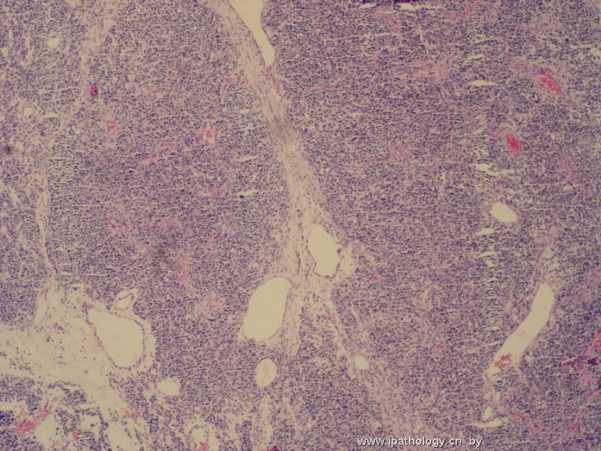
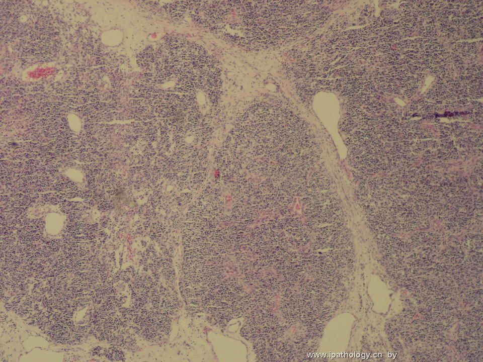
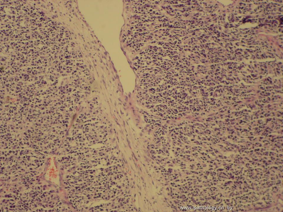
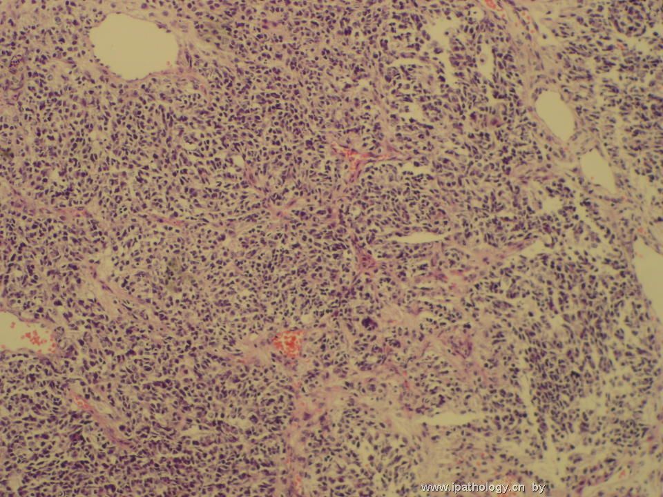
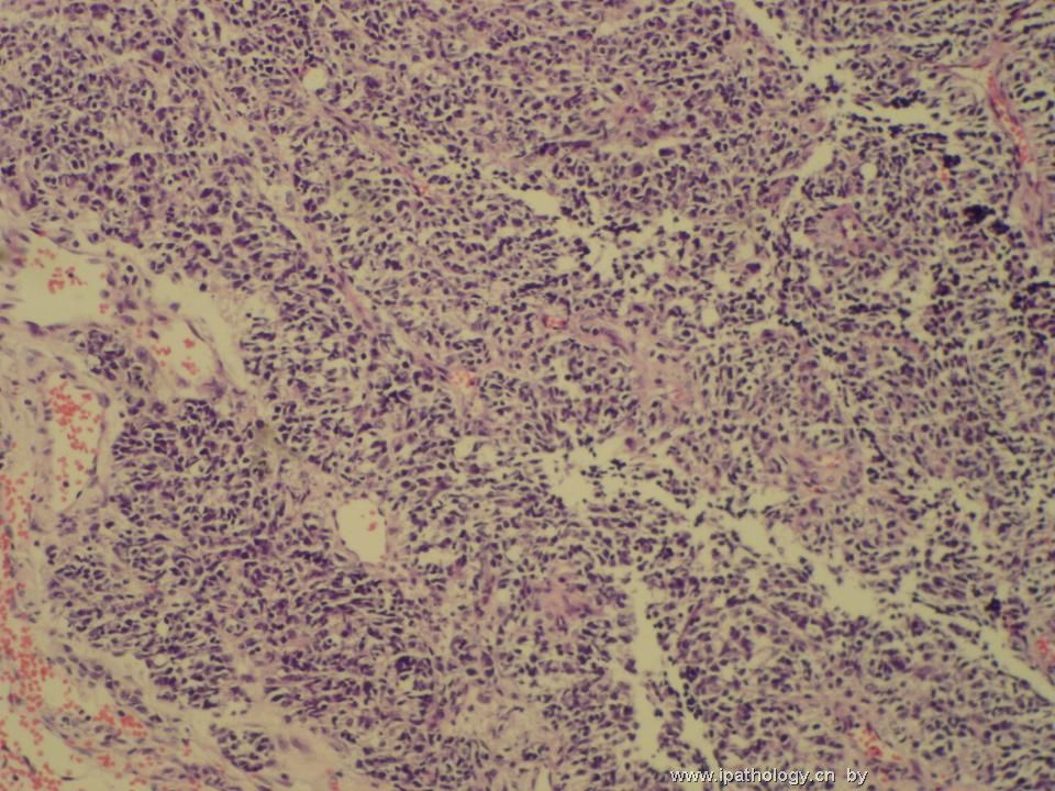
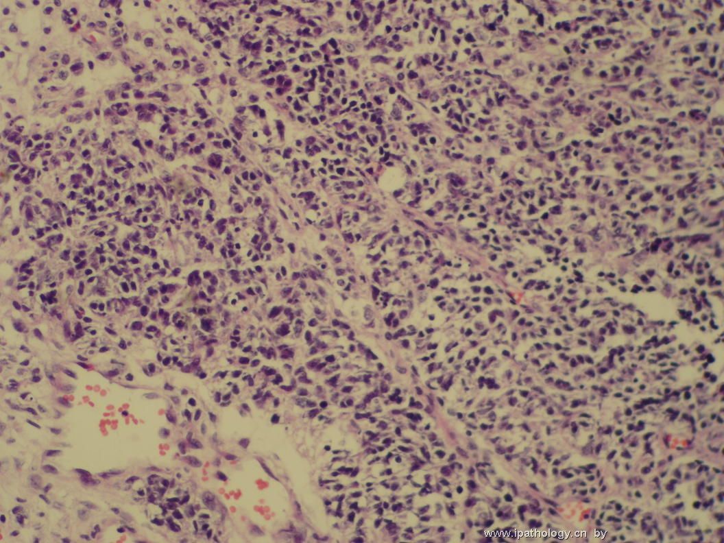
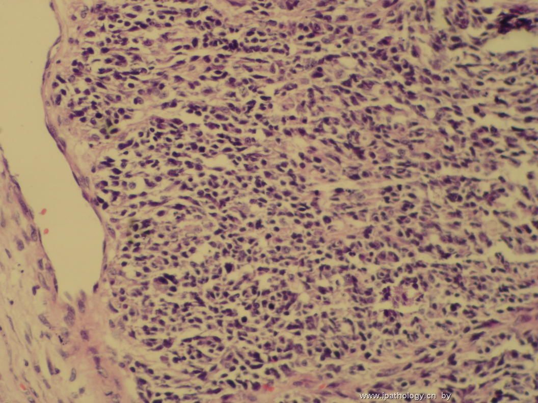
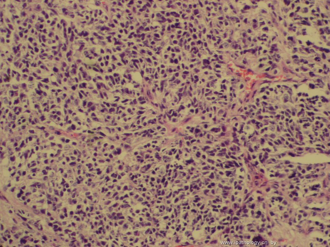
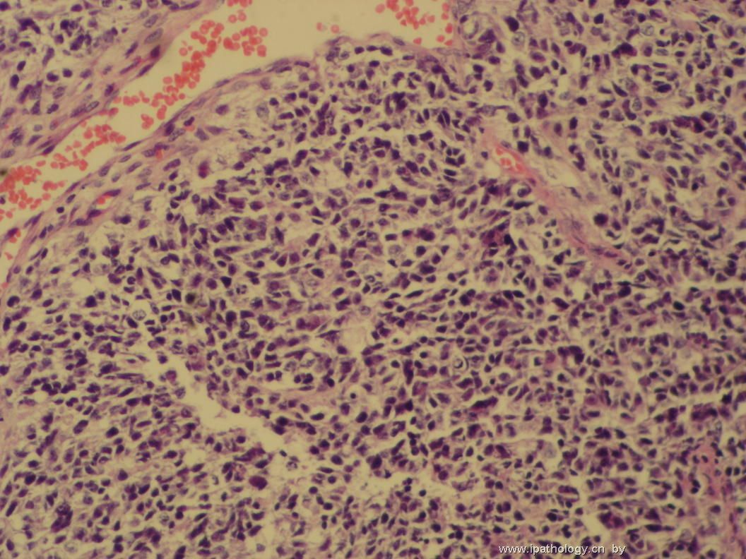
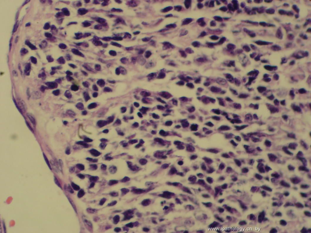
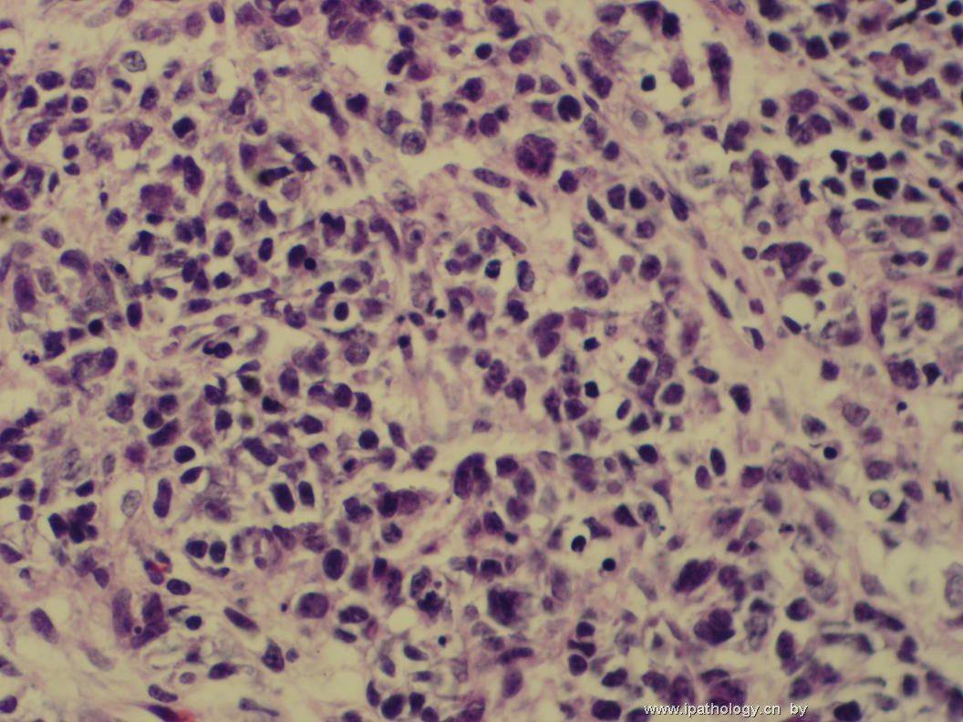
The low-power photos and anatomic location suggest hemangiopericytoma. However, the vascular pattern is not consistent with this possibility. High-power photos seem to show a few plasma cells and small lymphocytes in addition to poorly differentiated neoplastic cells. These neoplastic cells are pleomorphic and they seem to have clear cytoplasm. To me, a vague nested pattern is present. I do not favor a lymphoma. I don't see meningothelial differentiation or true rhabdoid cells to support rhabdoid meningioma. Dura-based PNET is rare, and should be carefully ruled out by immunohistochemical stains (GFAP, synaptophysin, NSE). Metastases certainly need to be ruled out. Is there any known malignancy (such as rhabdomyosarcoma, Ewing sarcoma, synovial sarcoma, paraganglioma, etc) in her clinical history? Please share your immunohistochemical results with us to narrow the search for the final diagnosis.

聞道有先後,術業有專攻
-
翻译:从低倍图像和解剖位置上看像血管外皮细胞瘤。但是血管结构不支持这种可能性。高倍图像在低分化肿瘤细胞之外可见少许浆细胞及小淋巴细胞。肿瘤细胞具有多形性,似乎有透明的胞浆。就我看来,有一个模糊的巢状结构。我不同意淋巴瘤。我没有看到脑膜瘤分化以及真正的横纹肌样细胞来支持横纹肌样脑膜瘤的诊断。硬脑膜发生的PNET很罕见,需要通过免疫组化染色(GFAP, synaptophysin, NSE)仔细地除外。转移瘤肯定也需要除外。在她的病史中是否有已知的恶性肿瘤?如横纹肌肉瘤、尤文瘤、滑膜肉瘤、副节瘤等等。请为我们提供免疫组化结果以集中搜寻正确答案的范围。

