| 图片: | |
|---|---|
| 名称: | |
| 描述: | |
- 右颞叶肿瘤
-
本帖最后由 于 2007-04-24 11:52:00 编辑
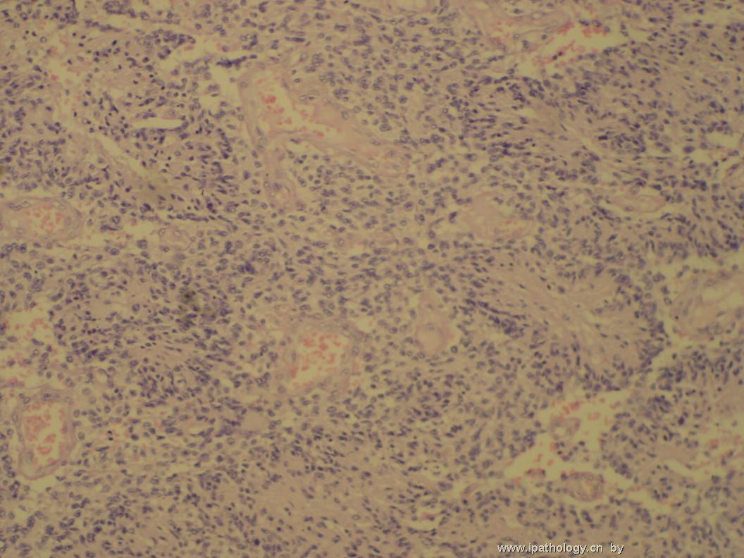
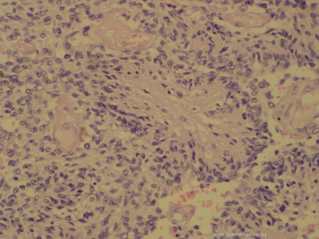
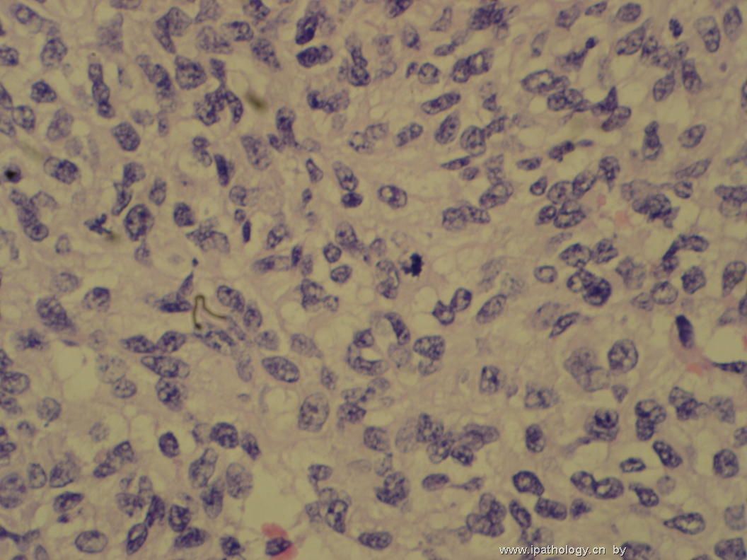
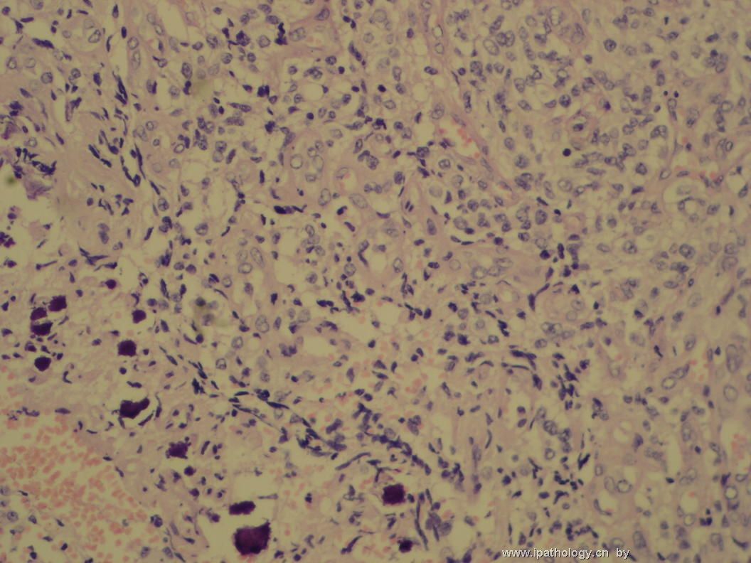
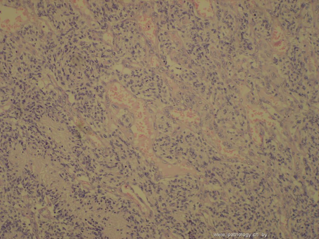
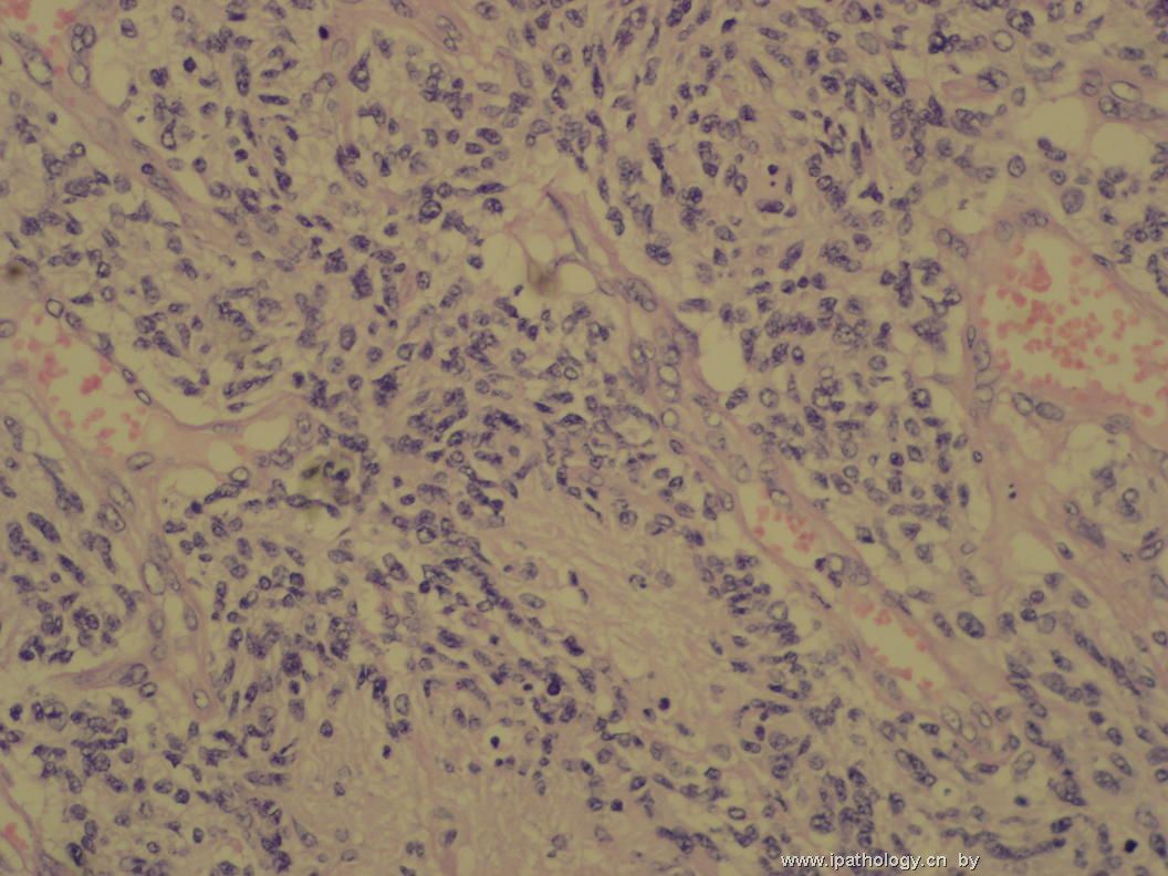
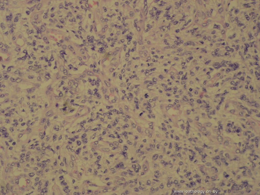
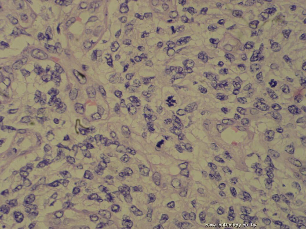
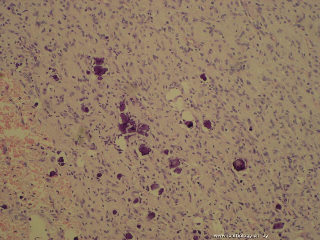
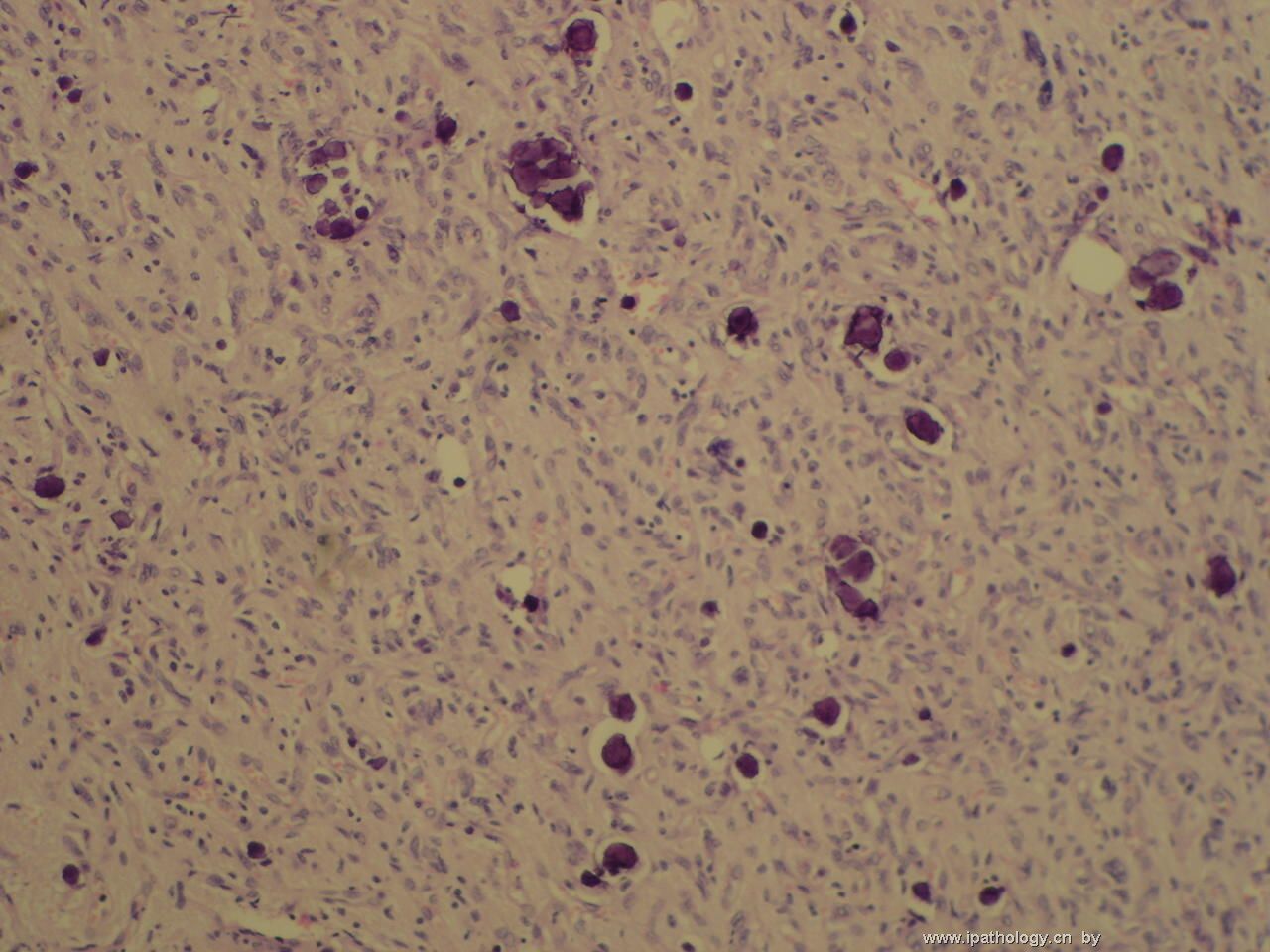
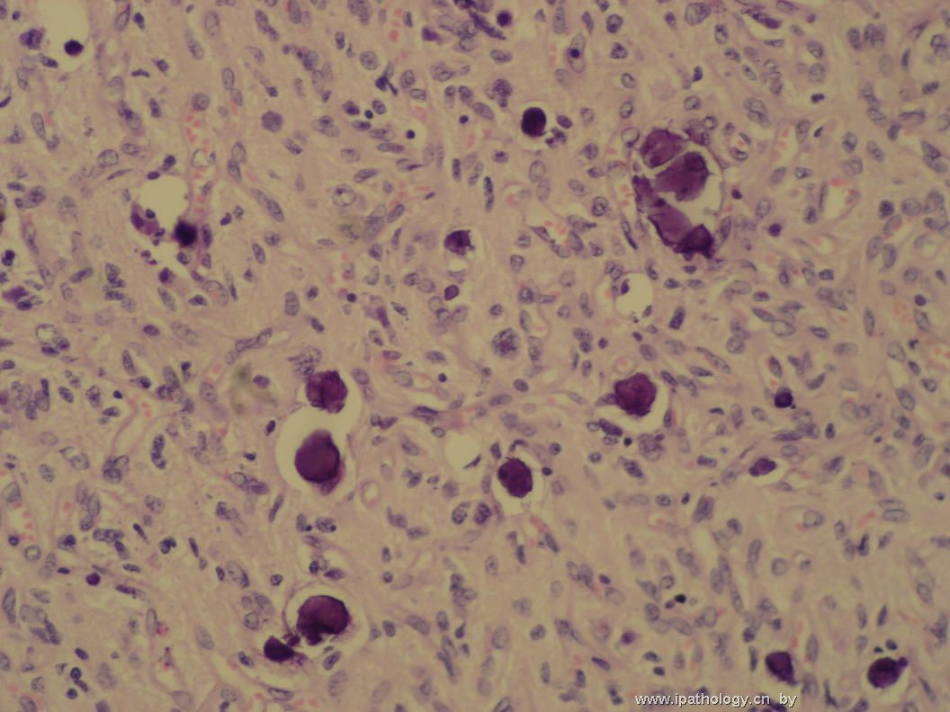
The blood vessels seen in this mitotically very active malignant neoplasm are not those encountered in meningioma. Instead, they remind me of neovasculature seen in high grade gliomas. I do not see meningothelial differentiation. The last two photos show calcospherites (they are not psammoma bodies) and associated neoplastic cells arranged in a pattern consistent with that seen in a low grade fibrillary astrocytoma. With neovasculature and easily identified mitoses in hypercellular areas, I suspect this is a small cell variant of WHO grade IV glioblastoma arising from a pre-existing low grade astrocytoma. The rare seemingly papillary structures may be found in meningioma (amianthoid fibers) as well as in some astrocytomas and mixed glioneuronal tumors. I do not see necrosis. GFAP and EMA immunohistochemical stains would help clarify the diagnosis.

聞道有先後,術業有專攻
-
翻译:在这个核分裂非常活跃的恶性肿瘤中见到的血管不是在脑膜瘤中可以遇到的那种。相反,这使我想起在高级别胶质瘤中可以见到的新生血管。我没有见到脑膜分化。最后两张照片显示钙盐沉积(它们不是砂砾体)以及相关的肿瘤细胞结构符合低级别纤维型星形细胞瘤。加上新生血管和在高细胞密度区域中易见的核分裂像,我怀疑这是一例自先前存在的低级别星形细胞瘤恶变而来的WHOIV级的胶母细胞瘤的一种小细胞变异。这些稀疏的表面上看起来的乳头状结构可以见于脑膜瘤(海绵样纤维)以及某些星形细胞瘤及神经元胶质混合肿瘤。我没有见到坏死。GFAP 和 EMA 染色可以帮助明确诊断。

