| 图片: | |
|---|---|
| 名称: | |
| 描述: | |
- educational case---rare gallbladder tumor
| 姓 名: | ××× | 性别: | female | 年龄: | 31 |
| 标本名称: | gallbladder | ||||
| 简要病史: | right abdominal pain | ||||
| 肉眼检查: | |||||
Patient presented to hospital with signs and symptoms of chronic cholecystitis and cholelithiasis. Ultrasound examination revealed thickening of gallbladder wall with
abnormal septation around liver bed. Patient underwent laparoscopic cholecystectomy and resection of the adjacent liver bed.
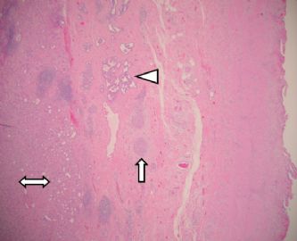
The gallbladder measures 9 × 4 × 3 cm and shows an indurated localized thickened fundic wall area measuring 2 ×1.5 × 1 cm located around the fundus and liver bed. Cut surface of the thickened area was off-white and firm. The gallbladder mucosa was normal. Three tan-yellow stones were present within gallbladder lumen. Gallbladder neck and cystic duct were grossly unremarkable.
-
本帖最后由 于 2009-04-26 21:06:00 编辑
| 以下是引用qianxun在2009-4-26 20:29:00的发言:
Patient presented to hospital with signs and symptoms of chronic cholecystitis and cholelithiasis. Ultrasound examination revealed thickening of gallbladder wall with abnormal septation around liver bed. Patient underwent laparoscopic cholecystectomy and resection of the adjacent liver bed.
The gallbladder measures 9 × 4 × 3 cm and shows an indurated localized thickened fundic wall area measuring 2 ×1.5 × 1 cm located around the fundus and liver bed. Cut surface of the thickened area was off-white and firm. The gallbladder mucosa was normal. Three tan-yellow stones were present within gallbladder lumen. Gallbladder neck and cystic duct were grossly unremarkable. | ||||||||||||||||||||||||
译文:部位 胆囊 临床症状:右侧腹痛
病人因慢性胆囊炎和胆囊结石而入院,超声检查发现胆囊壁增厚,胆囊与肝床之间有异常分隔。病人行腹腔镜下胆囊切除术和邻近肝床的切除
胆囊大小9 × 4 × 3 cm,胆囊底部有一个局限性硬结样的增厚,大小2 ×1.5 × 1 cm 。切面灰白色,质地致密。胆囊粘膜正常。胆囊腔内见有3个褐黄色石头。胆囊颈部和胆囊管大体上未见异常。
-
zhang197510 离线
- 帖子:409
- 粉蓝豆:2971
- 经验:448
- 注册时间:2009-03-22
- 加关注 | 发消息
-
liguoxia71 离线
- 帖子:4174
- 粉蓝豆:3122
- 经验:4677
- 注册时间:2007-04-01
- 加关注 | 发消息


