| 图片: | |
|---|---|
| 名称: | |
| 描述: | |
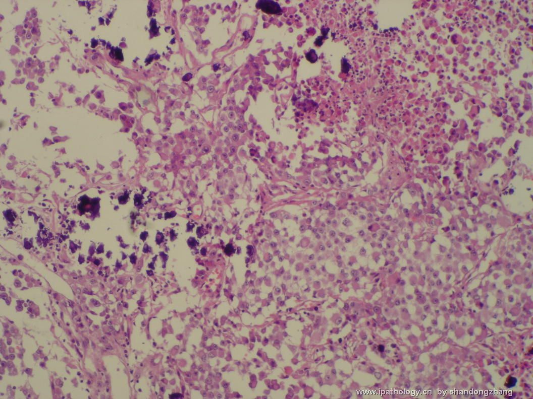
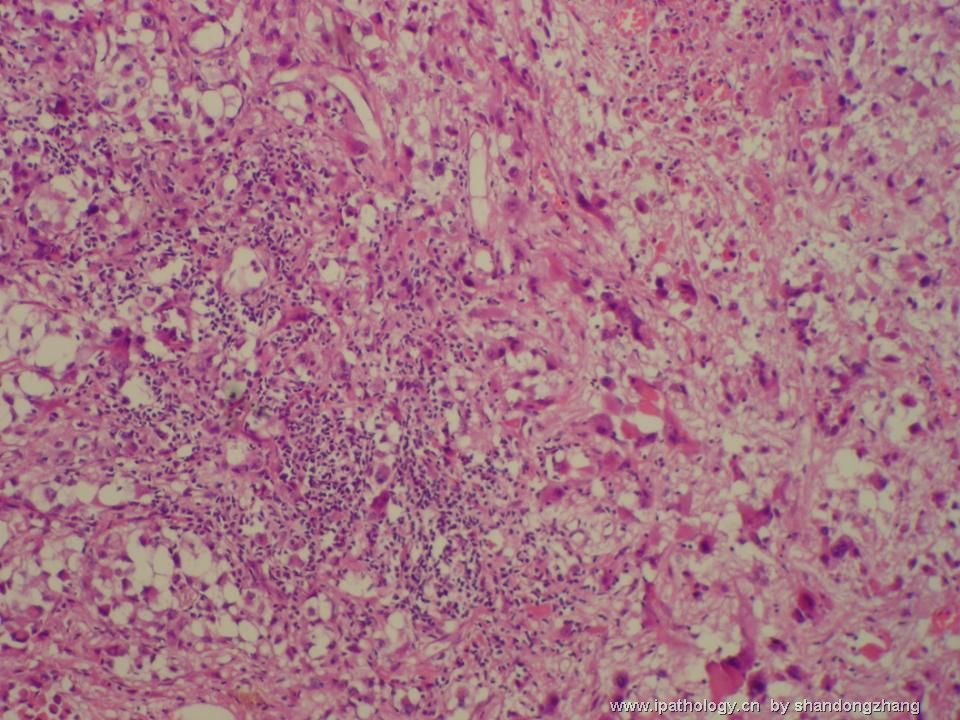
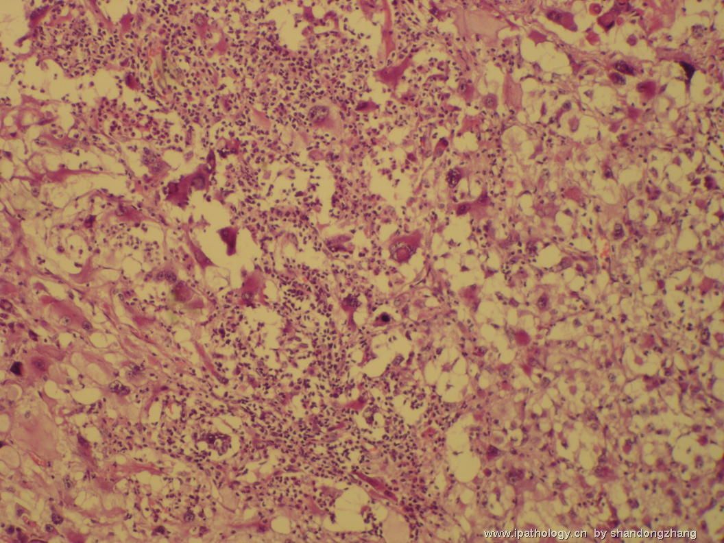
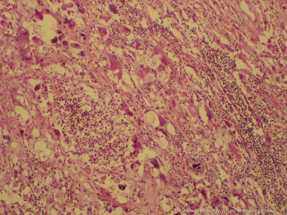
- 左颞叶肿瘤
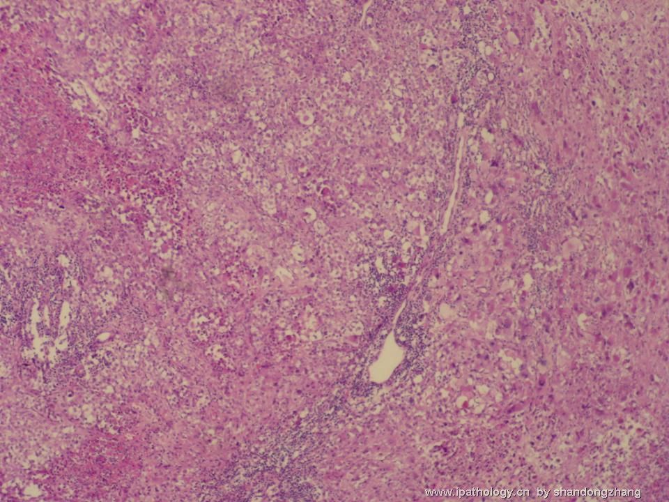
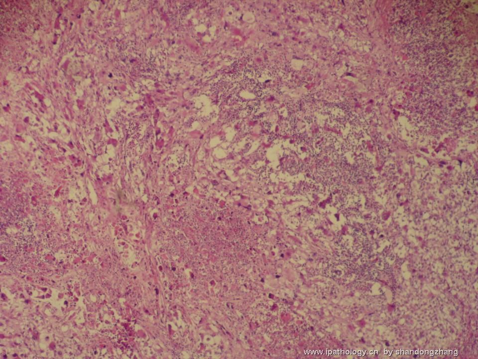
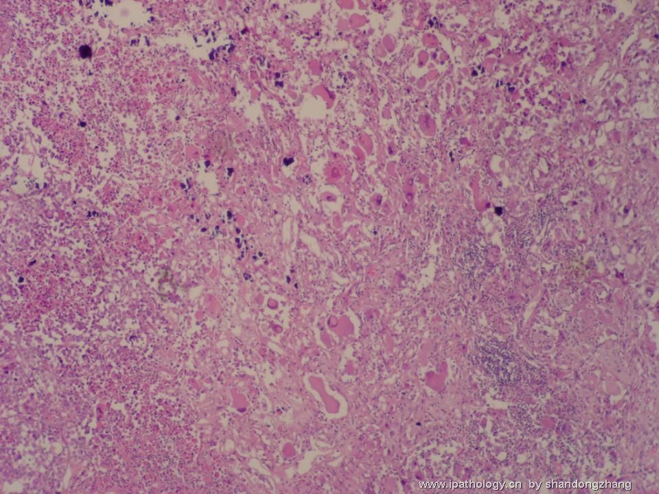
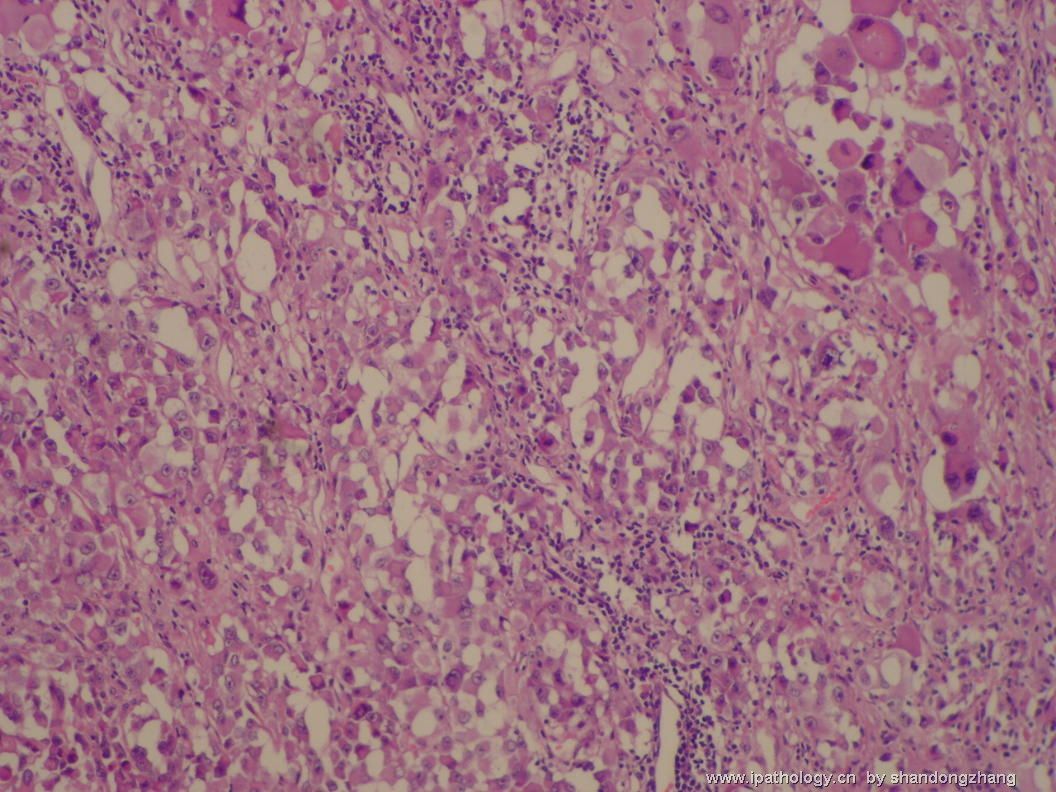
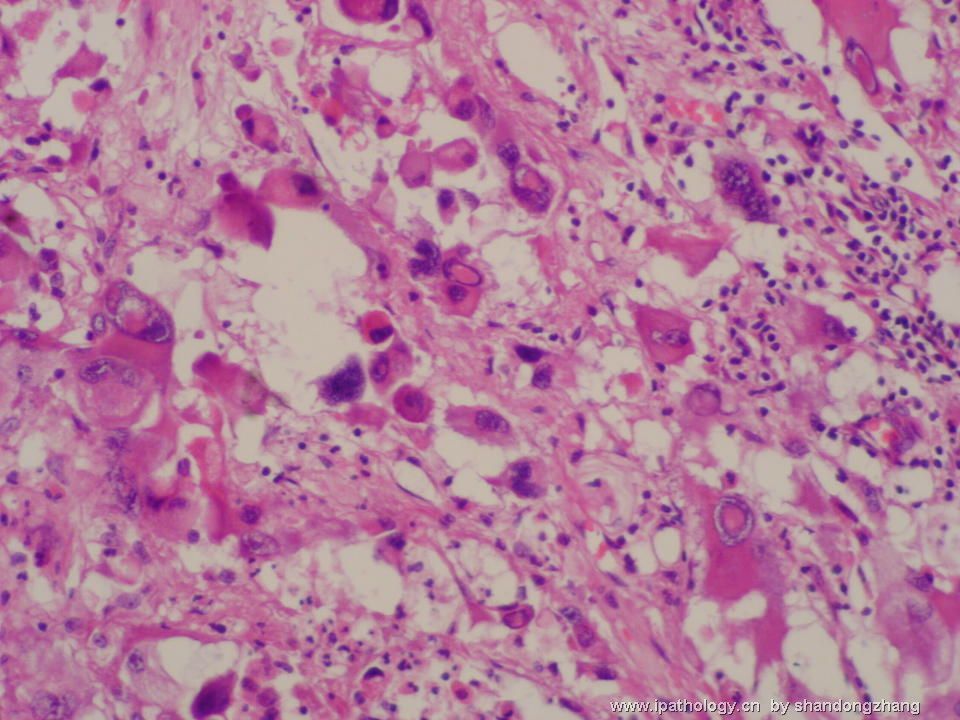
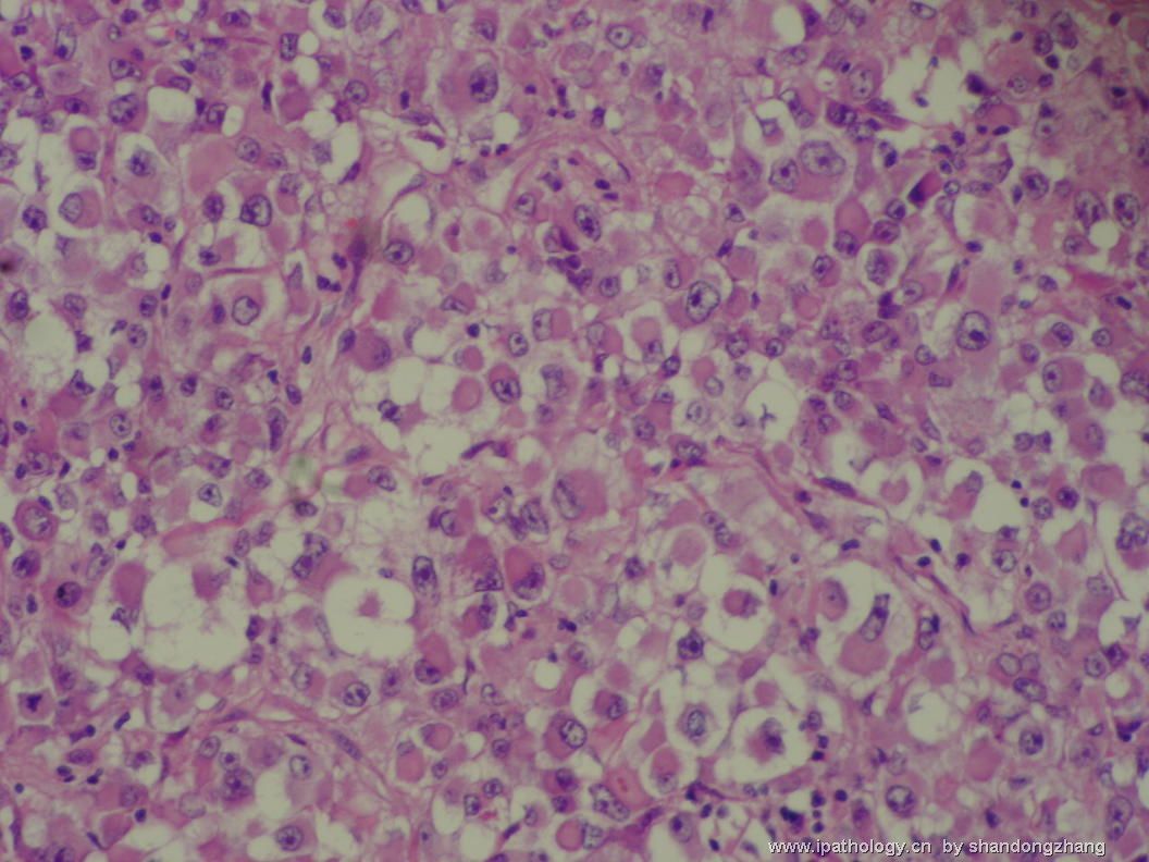
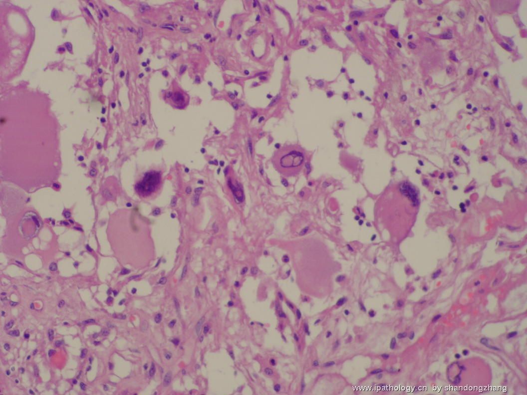
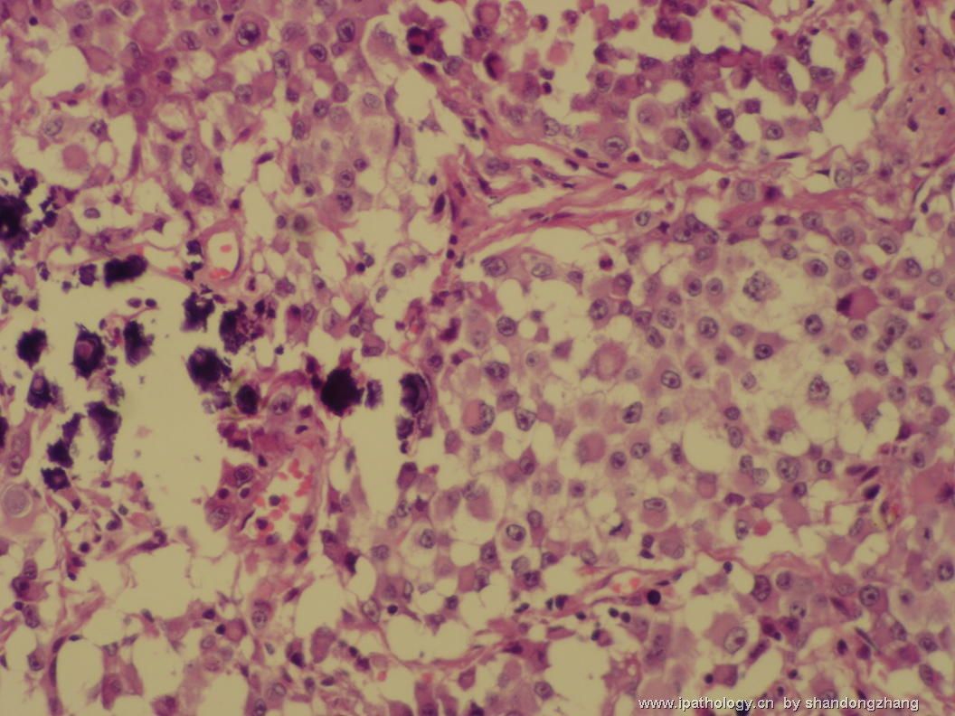
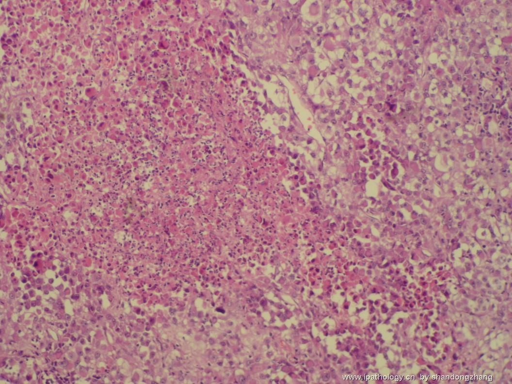
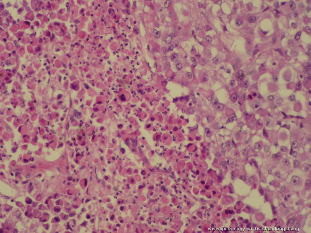
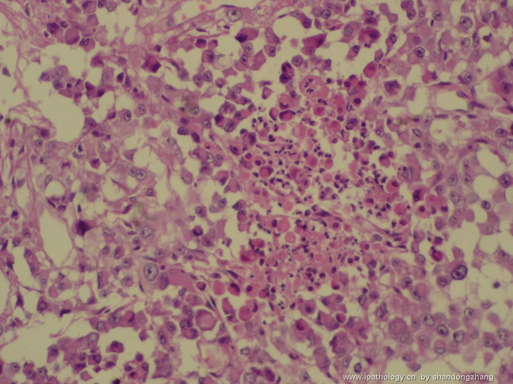
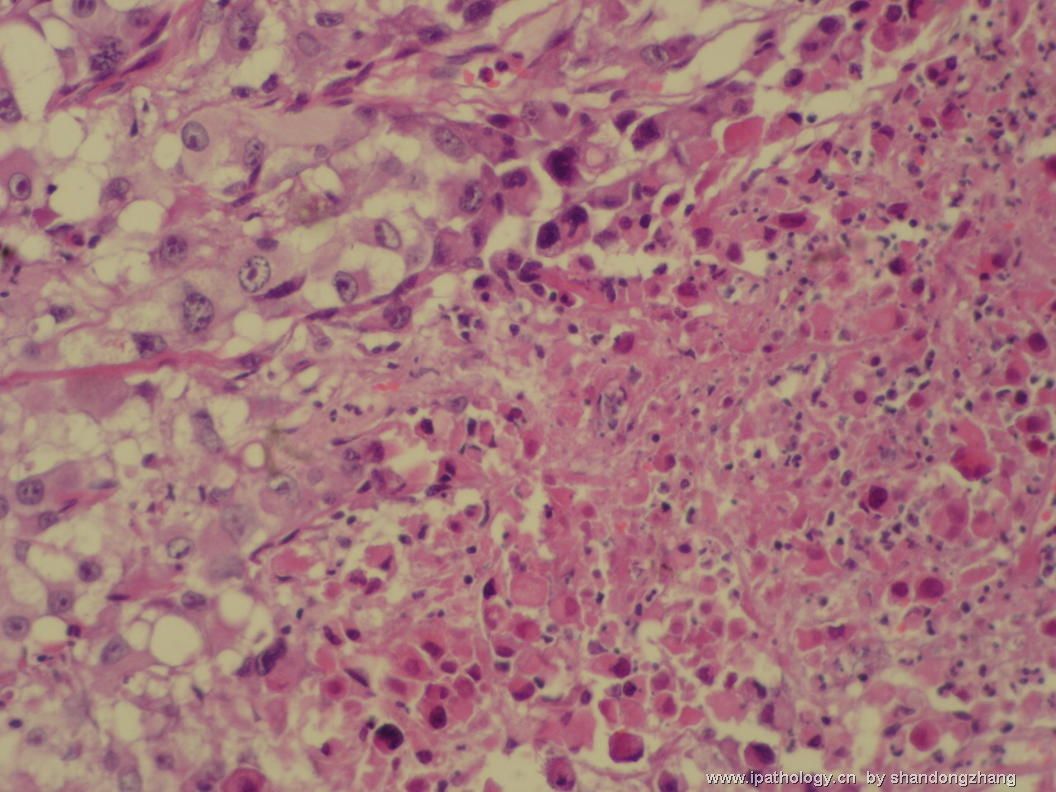
I raise the possibility of pleomorphic xanthoastrocytoma (PXA) because of the presence of inflammatory cells and large atypical giant cells with intranuclear pseudoinclusions and eosinophilic or vacuolated cytoplasm. Calcification and patient's age are both consistent with AT/RT and PXA. AT/RT. MRI images of these lesions are very different - PXA is circumscribed, focally cystic with an enhancing nodule, whereas AT/RT is massive with heterogeneous enhancement, irregularly shape and infiltration. I would look for eosinophilic granular bodies carefully.

聞道有先後,術業有專攻
-
wangzhen_01 离线
- 帖子:197
- 粉蓝豆:3
- 经验:197
- 注册时间:2006-10-04
- 加关注 | 发消息
-
ZQH19811029 离线
- 帖子:458
- 粉蓝豆:1
- 经验:458
- 注册时间:2009-11-15
- 加关注 | 发消息

