| 图片: | |
|---|---|
| 名称: | |
| 描述: | |
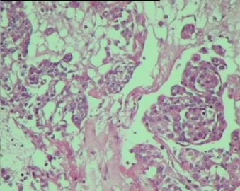
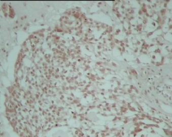
- 左侧额叶肿物
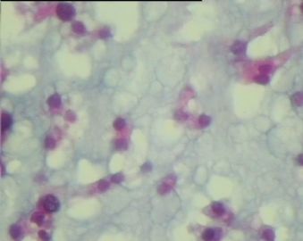
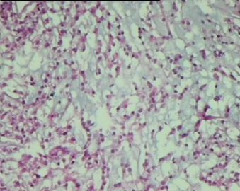
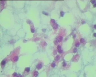
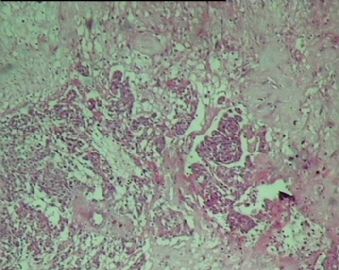
It is possible that this is a chordoid meningioma, especially if this is a frontal lobe lesion and not near the skull base midline. If the last photo is indeed stain for progesterone receptor protein, the diagnosis would be confirmed. Chordoid meningioma usually shows chordoid features only focally, and meningothelial whorls can be seen elsewhere in the same tumor. Chordoid chondrosarcoma and chordoma both can have areas looking exactly the same as this case (HE), and only immunohistochemical stains can help distinguish them:
Chordoid meningioma - EMA(+), PR(+), AE1(-), S100(+/-)
Chordoid chondrosarcoma - EMA(-), PR(-), AE1(-), S100(+)
Chordoma - EMA(+), PR(-), AE1(+), S100(-)
Another chordoid tumor in CNS is chordoid glioma of third ventricle. It is limited to this anatomic location and usually shows associated inflammation with strong GFAP immunoreactivity in all neoplastic cells.

聞道有先後,術業有專攻
-
zhang197510 离线
- 帖子:409
- 粉蓝豆:2971
- 经验:448
- 注册时间:2009-03-22
- 加关注 | 发消息
-
liangjinjun 离线
- 帖子:2328
- 粉蓝豆:2
- 经验:2457
- 注册时间:2007-08-07
- 加关注 | 发消息

| 以下是引用mjma在2009-4-5 7:32:00的发言:
It is possible that this is a chordoid meningioma, especially if this is a frontal lobe lesion and not near the skull base midline. If the last photo is indeed stain for progesterone receptor protein, the diagnosis would be confirmed. Chordoid meningioma usually shows chordoid features only focally, and meningothelial whorls can be seen elsewhere in the same tumor. Chordoid chondrosarcoma and chordoma both can have areas looking exactly the same as this case (HE), and only immunohistochemical stains can help distinguish them: Chordoid meningioma - EMA(+), PR(+), AE1(-), S100(+/-) Chordoid chondrosarcoma - EMA(-), PR(-), AE1(-), S100(+) Chordoma - EMA(+), PR(-), AE1(+), S100(-) Another chordoid tumor in CNS is chordoid glioma of third ventricle. It is limited to this anatomic location and usually shows associated inflammation with strong GFAP immunoreactivity in all neoplastic cells. |

