| 图片: | |
|---|---|
| 名称: | |
| 描述: | |
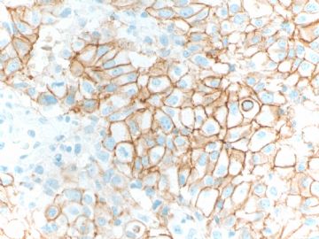
- B1580what scores of the IHC Her 2stain you will give (cqz 14)
Abin:
I read your last sentence: 流汗!我怎么看反了? I think again for your reaction. I will be very happy if you wrote that Zhao or Dr. Zhao, you were wrong in the score or why the score was 3 in the first case. We, Chinese are very traditionl. We respect olders, seninors, professors, experts, teachers, or authority. But in filelds of sciences, in medicine, especially in the internet, we should observe the truth. I am sure that you knew I was not correct (you maight think Zhao gave a stupid answer), but you still said 我怎么看反了.
You should say that zhao, 你怎么看反了.
Her2 FISH was performed on both cases.
Case 1: Fish ratio: 1.06
Case 2: Fish ratio: 2.08
Fish ratio=Her2/CRP17. CEP 17 is a centromeric probe for chromosome 17, where Her2 gene resides.
Some ones please explain above FISH results about Her 2 gene amplification, negative, equivocal, or positive for both cases.
Thanks, cz
| 以下是引用cqzhao在2009-3-12 7:16:00的发言:
Abin: I read your last sentence: 流汗!我怎么看反了? I think again for your reaction. I will be very happy if you wrote that Zhao or Dr. Zhao, you were wrong in the score or why the score was 3 in the first case. We, Chinese are very traditionl. We respect olders, seninors, professors, experts, teachers, or authority. But in filelds of sciences, in medicine, especially in the internet, we should observe the truth. I am sure that you knew I was not correct (you maight think Zhao gave a stupid answer), but you still said 我怎么看反了. You should say that zhao, 你怎么看反了. |
abin:我读了你最后一句“流汗!我怎么看反了?”。我再次思考了你的反应。如果你写“Zhao or Dr. Zhao,你评分错了,或者第一例为什么是3分”,我会很高兴。我们中国人很传统。我们尊敬长者、前辈、教授、专家、教授或者权威。但在科学领域,在医学,特别是在网上,我们应该观察真理。我确信你知道我错了(你可能想Zhao给了愚蠢的答案),但你仍然说自己怎么看反了。
你应该说,Zhao,你怎么看反了。
(abin回复:Sorry to Dr.Zhao,我虽然有过疑惑,但是并没有足够的自信。谢谢您的批评,网上的专业讨论应该毫无保留)

华夏病理/粉蓝医疗
为基层医院病理科提供全面解决方案,
努力让人人享有便捷准确可靠的病理诊断服务。
| 以下是引用cqzhao在2009-3-14 1:46:00的发言:
Her2 FISH was performed on both cases. Case 1: Fish ratio: 1.06 Case 2: Fish ratio: 2.08 Fish ratio=Her2/CRP17. CEP 17 is a centromeric probe for chromosome 17, where Her2 gene resides. Some ones please explain above FISH results about Her 2 gene amplification, negative, equivocal, or positive for both cases. Thanks, cz |

华夏病理/粉蓝医疗
为基层医院病理科提供全面解决方案,
努力让人人享有便捷准确可靠的病理诊断服务。
Prognostic Biomarkers - HER2/neu by FISH
Indications for use
The proto-oncogene HER-2/neu (c-erbB-2) resides on chromosome 17q and encodes a trans-membrane tyrosine kinase growth factor receptor. Amplification of the HER-2/neu gene, or overexpression of the HER-2/neu protein, is found in 20-30% of breast cancers. There is a greater than 90% correlation between gene amplification and protein overexpression. Some studies suggest that HER-2 gene amplification assessed by Fluorescent In Situ Hybridization (FISH) may improve the predictive ability of this marker for Trastuzumab (Herceptin®) therapies (1, 2), especially in the 10-20% of cases with equivocal (i.e. 2+) results for protein expression by IHC . The primary indication for assessing HER-2 by FISH today is in cases with equivocal IHC results (3, 4).
Scoring/interpretation
In our laboratory, we use PathVysion, a FDA-approved kit for HER-2 testing by FISH and follow the scoring and interpretation recommended by the manufacturer. In this procedure, the chromosome 17 centromere is marked with a green florescent signal and HER-2 gene with an orange florescent signal. Briefly, we assess 60 non-overlapping tumor cell nuclei and count the number of green and orange signals in each cell. Then, the overall gene-to-chromosome (17) ratio is calculated. A tumor is designated as “positive” for gene amplification if gene-to-chromosome (17) ratio is >2.0

Detailed procedure
We use FDA approved PathVysion kit to assess HER-2 by FISH. As per manufacturers’ recommendation, 20 tumor non-overlapping cell nuclei are enumerated to obtain the HER-2/chromosome 17 ratio.
Her2 gene is located in Chromosome 17.
CEP 17 is a centromeric probe for chromosome 17.
Now American Society of Clinical Oncology/College of American Pathologists Guideline:
Her2/CEP 17 ratio>2.2: positive for Her2 gene amplification.
Her2/CEP 17 ratio <1.8: negative for her2 gene amplification.
Her2/CEP 17 ration 1.8-2.2: Equivocal Her2 gene amplification.
If the ratio >=2.0, the patients are eligible for adjuvant trastuzumb treatment.
http://arpa.allenpress.com/pdf/i1543-2165-131-1-18.pdf
Above is American Society of Clinical Oncology/College of American Pathologists Guideline recommendations for human epidermal growth factor receptor 2 (Her2) testing in breast cancer.
This is the best and standard paper in the US.
To Abin:
In fact most American general pathologists do not know the information in details also in this area. I know some because I am a breast pathologist.
-
Basically I complete this topic. Anti Her 2 drug, Trastuzumab is too expensive for individual usage. Her2 issue may be not very mportant for general pathologists in China now. I assume it will become important soon.
Trastuzumab (Herceptin) is a monoclonal antibody that interferes with the HER2/neu receptor.
Thank all people for reading or discussing this topic.
cz
| 以下是引用cqzhao在2009-3-4 0:33:00的发言:
See this topic. http://www.ipathology.cn/forum/forum_display.asp?keyno=16236 The guidline paper. http://jop.ascopubs.org/cgi/content/full/3/1/48
Brief description about the scores Tissue fixed using formalin 8-96 hours. 0 = No staing is observed or membrane staining is observed in less than 10% of the tumor cells. 1+ = A faint/barely perceptoble membrane staining is detected in more than 10% of the tumor cells. The cells are only stained in part of their membrane score 0, 1 called negative 2+ = A weak to moderate complete membrane staining is observed in more than 10% of the tumor cells. 2+ means equivocal 3+ = A strongly complete membrane staining is observed in more than 30% of the tumor cells. 3+ means positive. 译: 关于评分的简要说明: 组织用福尔马林固定8-96小时。(10%中性福尔马林?) 0=没有染色或少于10%的瘤细胞的细胞膜染色。 1+=10%以上的瘤细胞有微弱染色,且染色仅限于部分细胞膜。 得0分和1分为阴性。 2+ =大于10%的瘤细胞的细胞膜有微弱到中等强度的完整染色。 2+为可疑阳性。 3+ =大于30%的瘤细胞的细胞膜有完整的高强度的染色。 3+为阳性。
|

- 我思故我在! know something about everything,know everything about something.
-
本帖最后由 于 2009-03-22 12:03:00 编辑
| 以下是引用cqzhao在2009-3-21 9:39:00的发言:
Prognostic Biomarkers - HER2/neu by FISHIndications for use Scoring/interpretation
Detailed procedure |
This part is feom Baylor College of Medicine website. http://www.breastcenter.tmc.edu/research/cores/path/services/her2_fish.htm
预后性生物指标--her2/neu的FISH检测
应用指征:
原癌基因her2/neu位于17号染色体长臂上,编码跨膜酪氨酸激酶生长因子受体。20%-30%的乳腺癌中发现有her2/neu基因的扩增或her2/neu蛋白的过度表达。基因扩增和蛋白过表达之间的关联度大于90%。有研究表明,用FISH来评估her2的扩增可以提高这种指标在赫赛汀治疗中的预测效能,尤其是在10%-20%的免疫组化标记的蛋白表达结果可疑阳性的病例中。现在FISH检测her2最主要的适应症就是那些免疫组化检测结果可疑阳性的病例。
评分和解释
在我们实验室,我们用FDA批准的一种her2的FISH检测试剂盒-Pathvysion,按照厂家的要求来进行评分和解释。在检测过程中,17号染色体的着丝点被绿色的荧光标记,her2被标记为开花状的橙色荧光。简言之,我们评价60个不重叠的肿瘤细胞核并每个细胞内的对绿色信号和橙色信号进行计数,然后计算全部的基因/17号染色体的比率,如果该比率大于2.0被看作基因扩增“阳性”。
详细的过程:
我们用FDA批准的pathvysion试剂盒来做her2的FISH,按照厂家的推荐,计数20个不重叠的肿瘤细胞来获得her2基因/17号染色体的比率。

- 我思故我在! know something about everything,know everything about something.
| 以下是引用cqzhao在2009-3-4 0:33:00的发言: …… Brief description about the scores Tissue fixed using formalin 8-96 hours. 0 = No staing is observed or membrane staining is observed in less than 10% of the tumor cells. 1+ = A faint/barely perceptoble membrane staining is detected in more than 10% of the tumor cells. The cells are only stained in part of their membrane score 0, 1 called negative 2+ = A weak to moderate complete membrane staining is observed in more than 10% of the tumor cells. 2+ means equivocal 3+ = A strongly complete membrane staining is observed in more than 30% of the tumor cells. 3+ means positive. |
我们应用的诊断标准和你们一样,但我们在判断3+时比较保守一些,怕吃官司。比如对于病例1和病例2我们都会判为2+。叫病人花几千块钱做FISH,总比花十几万块钱做无效治疗强。还不会被告上法庭。(况且国内免疫组化的质量控制还存在一些问题)

- 博学之,审问之,慎思之,明辨之,笃行之。
笃行者 said: 我们应用的诊断标准和你们一样,但我们在判断3+时比较保守一些,怕吃官司。比如对于病例1和病例2我们都会判为2+。叫病人花几千块钱做FISH,总比花十几万块钱做无效治疗强。还不会被告上法庭。(况且国内免疫组化的质量控制还存在一些问题)
This is very good. I used the same pholosophy for the evaluation of Her2 IHC. Until the stains are very strong like positive control (see my above control photos), otherwise we call 2+ and the FISH will be done.
In fact I called 2+ for both the case 1 and 2. This is why I can have the FISH result.
Learning to evaluate Her2 IHC stains is very important. Hope all pathologists are aware of this espcically for the people who do not read the stains often.


