| 图片: | |
|---|---|
| 名称: | |
| 描述: | |
- 颅内肿物,胶质瘤?
-
liangjinjun 离线
- 帖子:2328
- 粉蓝豆:2
- 经验:2457
- 注册时间:2007-08-07
- 加关注 | 发消息
-
zhang197510 离线
- 帖子:409
- 粉蓝豆:2971
- 经验:448
- 注册时间:2009-03-22
- 加关注 | 发消息
-
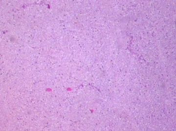
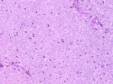
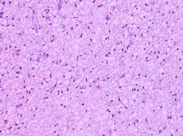
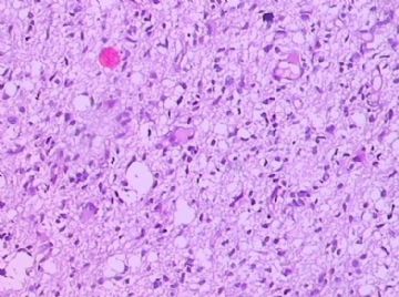
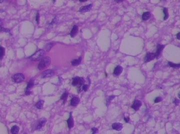
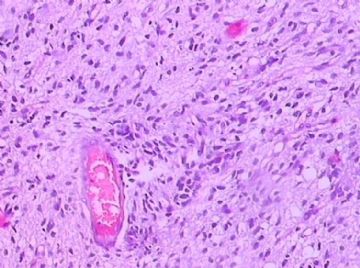
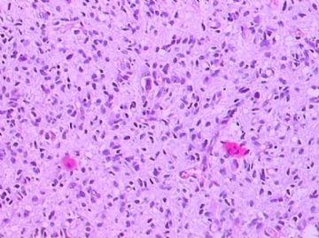
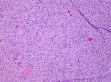
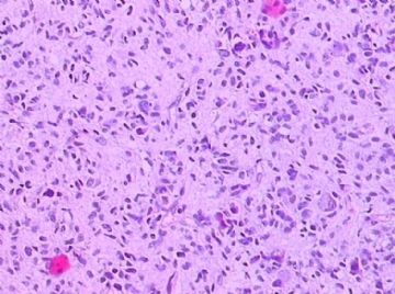
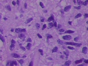
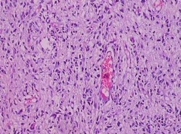
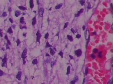
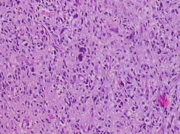
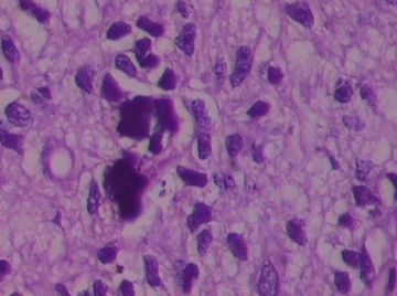
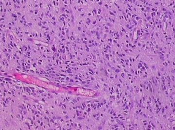
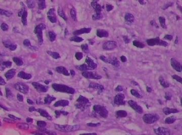
As many who have commented, I agree with the diagnosis of fibrillary astrocytoma. The grade of this neoplasm is difficult to decide based on the photos. There is calcification to suggest a low grade (WHO grade II) diffuse astrocytoma, but cellularity is high focally and nuclear atypia significant. I have not seen a mitotic figure in any of the photos. Please do a careful count of mitotic figures and look at the pre-surgical MRI of the lesion. If very rare mitoses are found (<3 per slide) and no contrast enhancement was detected on MRI, I would favor WHO grade II diffuse astrocytoma. On the other hand, if mitotic figures are easily seen (either focally or diffusely) and there was MRI contrast enhancement, I would favor WHO grade III anaplastic astrocytoma.

聞道有先後,術業有專攻
-
zhongshihua 离线
- 帖子:1608
- 粉蓝豆:0
- 经验:1651
- 注册时间:2006-09-11
- 加关注 | 发消息

