| 图片: | |
|---|---|
| 名称: | |
| 描述: | |
- 脑复发肿瘤-曾在北京知名医院诊断为间变性少突胶质细胞瘤
-
lanyueliang 离线
- 帖子:679
- 粉蓝豆:11
- 经验:1424
- 注册时间:2008-11-12
- 加关注 | 发消息
-
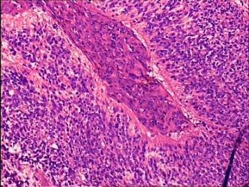
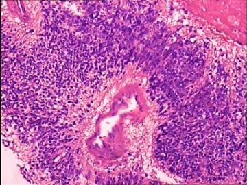
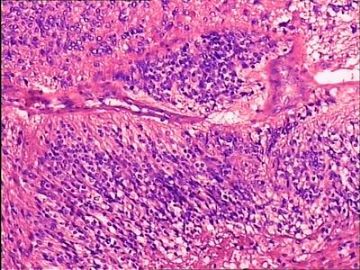
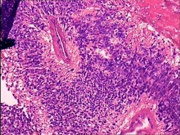
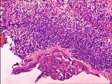
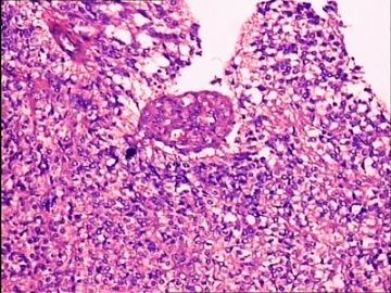
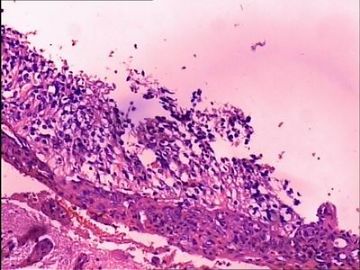
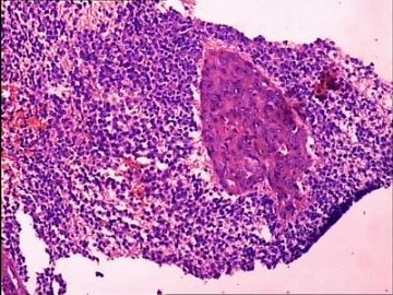
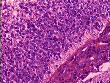
The high cellularity, vascular proliferation, and high MIB-1 labeling index are that of a malignancy. Neoplastic cells are relatively small with either elongated or round to slightly oval nuclei, uniform nuclear shape and size (at least focally) and little cytoplasm. GFAP was reported as negative, which would be unusual for anaplastic ependymoma, anaplastic oligodendroglioma/mixed oligoastrocytoma, and glioblastoma. There is a past history of anaplastic oligodendroglioma, and I believe that limits your differential diagnoses to anaplastic oligodendroglioma and glioblastoma with an oligodendroglioma component (the two are not much different in histopathology). To be sure, I would stain for EMA to rule out anaplastic ependymoma, and synaptophysin and NSE to rule out primitive neuroectodermal tumor (PNET). A challenging case this is.

聞道有先後,術業有專攻

