| 图片: | |
|---|---|
| 名称: | |
| 描述: | |
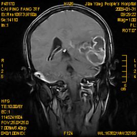
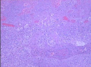
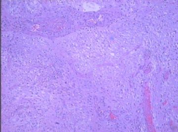
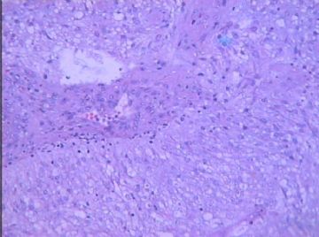
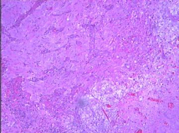
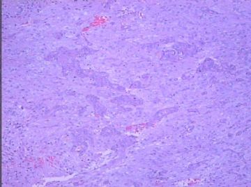
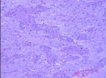
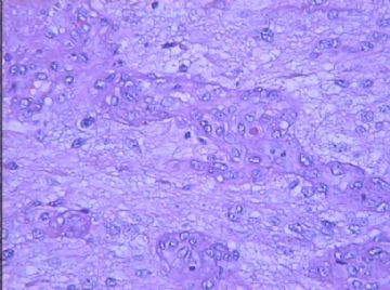
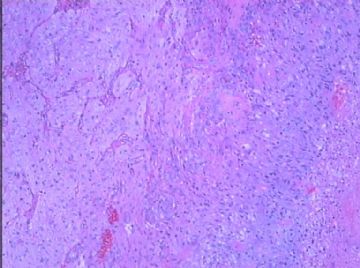
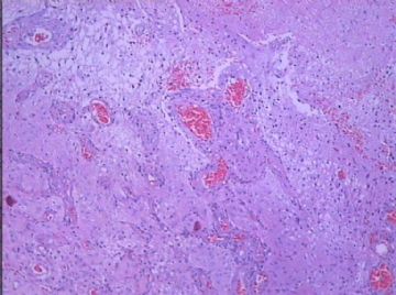
- 混合型少突胶质星形细胞瘤(WHO3级)?
-
zhang197510 离线
- 帖子:409
- 粉蓝豆:2971
- 经验:448
- 注册时间:2009-03-22
- 加关注 | 发消息
-
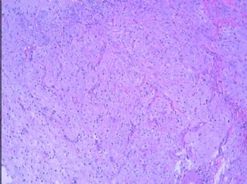
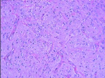
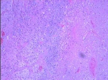
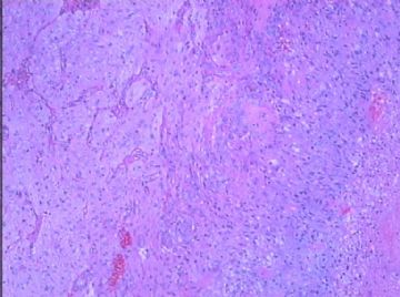
The MRI image is very helpful. The photomicrographs are somewhat blue in background, but I could see focal tumor necrosis and vascularp proliferation. There are elongated neoplastic astrocytes, but the overall cellularity is not very high in the photos. Could you tell whether mitotic figures are found easily? And whether there are hypercellular areas not shown in the photos? More importantly, are there dysplastic neurons in the neoplasm? My suspicion is that this is WHO grade IV glioblastoma, but I would like to rule out a ganglioglioma because of low cellularity.

聞道有先後,術業有專攻
-
liangjinjun 离线
- 帖子:2328
- 粉蓝豆:2
- 经验:2457
- 注册时间:2007-08-07
- 加关注 | 发消息

